


Perineural Invasion Is Associated With Stromal Remodeling, Angiogenesis, and Neural Features in Colorectal Cancer
DOI:
https://doi.org/10.14740/wjon2889Keywords:
Colorectal cancer, Perineural invasion, Tumor microenvironment, Stromal remodeling, CMS4, Angiogenesis, Neural developmentAbstract
Background: Perineural invasion (PNI) is known as a pathological indicator of aggressive tumor behavior and is associated with poor survival in colorectal cancer (CRC). Although individual biological features associated with PNI have been reported, the relationship between PNI and neural cell attraction and stromal features within the tumor microenvironment remains incompletely understood. We aimed to characterize the clinical, genomic, immune, stromal, vascular, and neural features associated with PNI-positive CRC.
Methods: Clinical and transcriptomic data from 179 CRC patients in The Cancer Genome Atlas (TCGA) were analyzed. PNI status was morphologically determined from pathological records, yidlding 45 PNI-positive and 134 PNI-negative cases. Gene set enrichment analysis (GSEA) using Hallmark gene sets characterized transcriptomic differences. Genomic features, immune cell composition, consensus molecular subtypes (CMS), stromal and vascular features, and neural features were compared between groups.
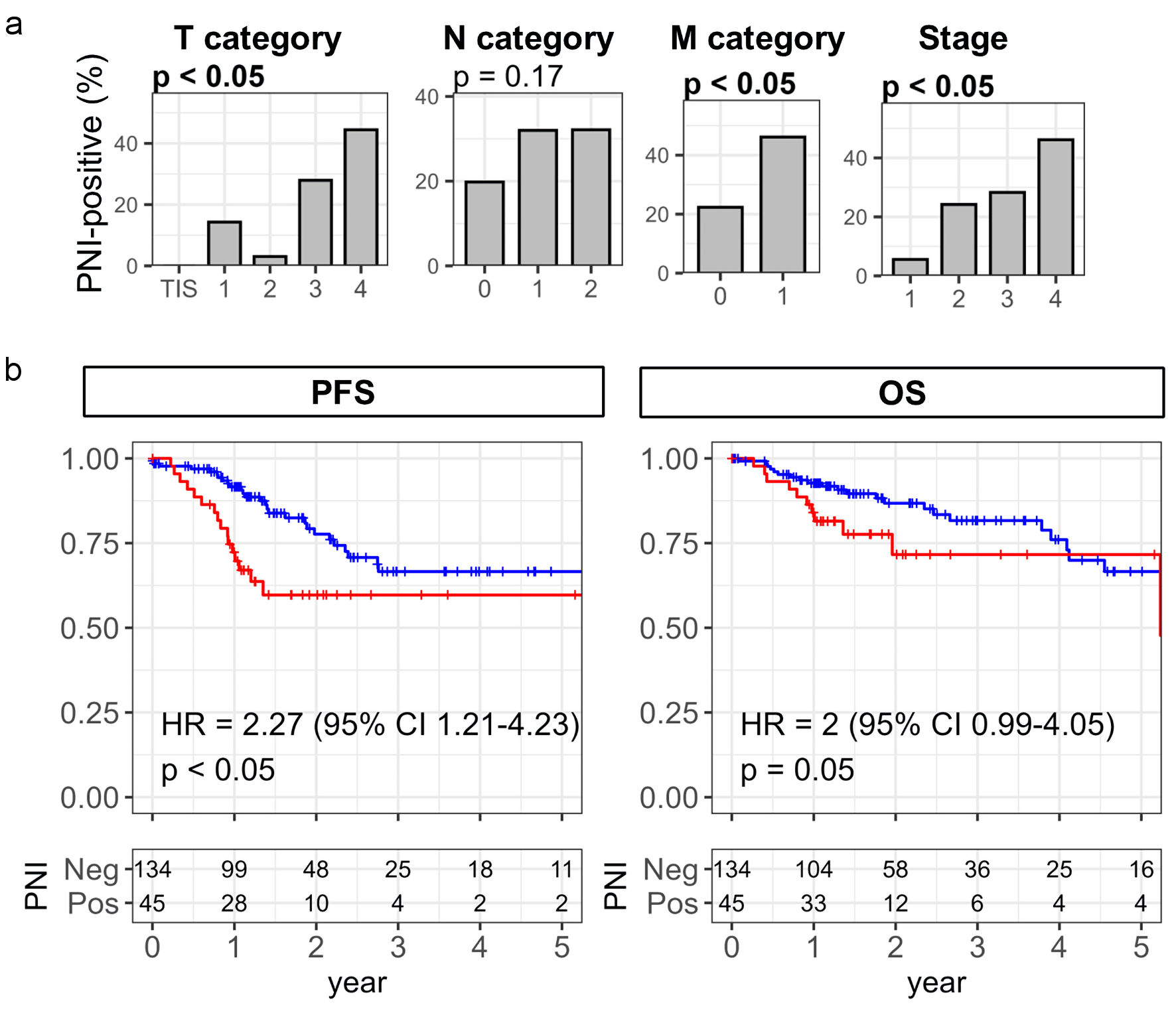
Results: PNI-positive CRC was associated with advanced T category, distant metastasis, advanced stage, and worse progression-free survival. There was the enrichment of proliferation-related pathways, including E2F targets, the G2M checkpoint and MYC target pathways, in PNI-positive CRC. However, genomic differences were limited, with higher homologous recombination deficiency and TP53 mutation frequency in PNI-positive CRC. PNI was not associated with broad changes in xCell immune scores but showed reduced interferon-α and interferon-γ responses. PNI-positive CRC was enriched in the mesenchymal consensus molecular subtype 4 (CMS4) and showed increased stromal fraction and extracellular matrix gene expression. Vascular features were also increased, including endothelial cells and lymphatic endothelial cells, together with enrichment of angiogenesis and increased transforming growth factor (TGF)-β-related features. Notably, PNI-positive CRC showed increased Schwann cell and neuron projection guidance scores and higher expression of axon guidance genes. These findings identified concurrent stromal, vascular, and neural features associated with PNI-positive CRC.
Conclusions: PNI-positive CRC is characterized by a coordinated mesenchymal and stromal phenotype accompanied by angiogenic and neural features, with relatively limited genomic and immune differences. These findings provide a biological framework that may help explain the aggressive clinical behavior of PNI-positive CRC.
Published
Issue
Section
License
Copyright (c) 2026 The authors

This work is licensed under a Creative Commons Attribution 4.0 International License.