Spatial Immune Remodeling Across the Gallbladder Carcinogenesis Spectrum: A Multicenter Digital Pathology Study
DOI:
https://doi.org/10.14740/wjon2771Keywords:
Gallbladder carcinoma, Tumor immune microenvironment, CD8, FOXP3, CD163, Tumor-associated macrophages, Immune exclusion, Digital pathology, Biliary intraepithelial neoplasiaAbstract
Background: Gallbladder carcinoma (GBC) is the most aggressive malignancy of the biliary tract and is commonly associated with late diagnosis and early metastatic dissemination. Although the morphological sequence of gallbladder carcinogenesis from chronic inflammation to epithelial dysplasia and invasive carcinoma is well recognized, the role of the tumor immune landscape during this process remains insufficiently characterized. This study aimed to quantitatively evaluate immune microenvironment alterations across the spectrum of gallbladder lesions using immunohistochemistry and digital pathology analysis.
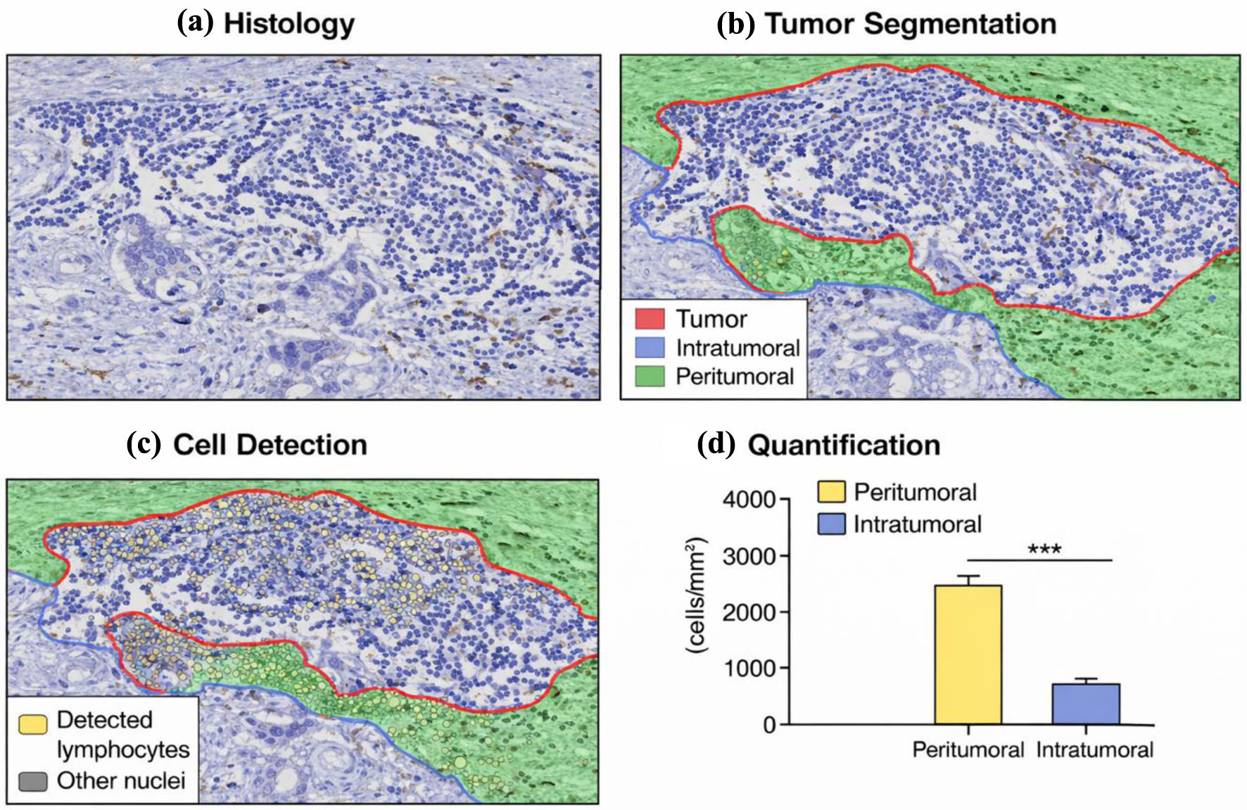
Methods: A retrospective multicenter study was conducted using formalin-fixed paraffin-embedded gallbladder specimens collected between 2015 and 2026 from several collaborating hospitals and analyzed at the Department of Molecular Pathology, Tbilisi State Medical University. The study cohort included 90 cases representing chronic cholecystitis, intestinal metaplasia, biliary intraepithelial neoplasia (BilIN-1, BilIN-2, BilIN-3), and invasive gallbladder adenocarcinoma. Immunohistochemical analysis was performed for CD8 (cytotoxic T lymphocytes), FOXP3 (regulatory T cells), and CD163 (tumor-associated macrophages). Whole-slide digital image analysis using QuPath and ImageJ was applied to quantify immune cell densities and evaluate spatial distribution between tumor center and invasive tumor front compartments. Composite immune indices were calculated to integrate cytotoxic and immunosuppressive immune populations. The immune suppression index was defined as (FOXP3 + CD163)/CD8.
Results: Quantitative analysis demonstrated progressive remodeling of the immune landscape across the spectrum of gallbladder lesions. CD8-positive cytotoxic lymphocytes were present in all lesion categories but showed heterogeneous density in invasive carcinoma. In contrast, FOXP3-positive regulatory T cells and CD163-positive macrophages demonstrated increased infiltration in dysplastic lesions and invasive tumors. Spatial analysis revealed preferential localization of CD8-positive lymphocytes at the invasive tumor margin with reduced infiltration of the tumor center, consistent with an immune exclusion pattern. Tumors associated with liver metastases exhibited lower cytotoxic lymphocyte density and increased infiltration by regulatory T cells and macrophages. Composite immune suppression indices were correspondingly elevated in metastatic tumors.
Conclusions: Gallbladder carcinogenesis is associated with significant remodeling of the tumor immune microenvironment characterized by enrichment of immunosuppressive immune populations and spatial restriction of cytotoxic lymphocyte infiltration. Digital pathology-based quantification of immune architecture provides reproducible assessment of tumor–immune interactions and may contribute to the identification of immune-related biomarkers of tumor aggressiveness in GBC.
Published
Issue
Section
License
Copyright (c) 2026 The authors

This work is licensed under a Creative Commons Attribution 4.0 International License.