Transcription Factor MAX Regulates Liver Cancer Cell Growth, Migration, Invasion, and Epithelial–Mesenchymal Transition by Promoting SF3A3 Expression
DOI:
https://doi.org/10.14740/wjon2783Keywords:
Hepatocellular carcinoma, MAX, SF3A3, Alternative splicing, Transcription factor, Cancer stemness, Therapeutic target, Epithelial–mesenchymal transitionAbstract
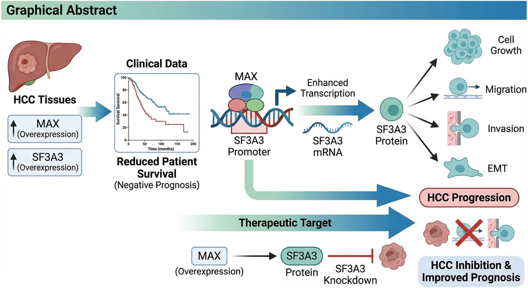
Background: The regulatory relationship between transcription factor MAX and splicing factor SF3A3 in hepatocellular carcinoma (HCC) is unknown. We investigated whether MAX directly regulates SF3A3 and their functional role in HCC progression.
Methods: MAX and SF3A3 expression were analyzed in The Cancer Genome Atlas Liver Hepatocellular Carcinoma (TCGA-HCC) dataset (UALCAN, GEPIA) and validated in 33 paired HCC and adjacent non-tumor tissues by quantitative reverse transcription polymerase chain reaction (qRT-PCR). Expression and prognostic significance were assessed across cancer stages, grades, and nodal status. Functional roles were evaluated in Hep3B (high expression) and PLC/PRF/5 (low expression) cells using shRNA-mediated knockdown and overexpression, respectively. Cell proliferation (cell counting kit-8, colony formation), migration (wound healing, Transwell), invasion (Matrigel Transwell), and epithelial–mesenchymal transition (EMT) (Western blot for E-cadherin, N-cadherin, and vimentin) were assessed. Mechanistic studies included chromatin immunoprecipitation (ChIP)-quantitative polymerase chain reaction (qPCR), luciferase reporter assays with site-directed mutagenesis, and Myc inhibition. In vivo tumor growth was evaluated using a xenograft mouse model.
Results: MAX and SF3A3 were overexpressed in HCC tissues compared to normal liver, and high expression was correlated with reduced patient survival, advanced cancer stage, higher tumor grade, and nodal metastasis. A significant positive correlation between MAX and SF3A3 expression was observed. Functional assays demonstrated that MAX or SF3A3 overexpression promoted HCC cell proliferation, migration, invasion, and EMT, while knockdown suppressed these phenotypes. MAX directly bound the SF3A3 promoter (P3 E-box) and activated its transcription in a Myc-dependent manner. Overexpression of MAX or SF3A3 promoted malignant phenotypes, while SF3A3 knockdown reversed MAX-driven oncogenic effects in vitro and reduced tumor growth in vivo.
Conclusion: This study establishes a novel MAX-SF3A3 regulatory axis in which MAX directly binds the SF3A3 promoter and activates its transcription in a Myc-dependent manner, driving HCC cell proliferation, migration, invasion, EMT, and tumor growth. Targeting the MAX-SF3A3 axis represents a potential therapeutic strategy for HCC.
Published
Issue
Section
License
Copyright (c) 2026 The authors

This work is licensed under a Creative Commons Attribution 4.0 International License.