Immunohistochemistry as a Cornerstone in Lung Cancer Diagnosis: Subtyping Based on the 2021 World Health Organization Classification and Its Practical Application
DOI:
https://doi.org/10.14740/wjon2757Keywords:
Lung carcinoma, Immunohistochemistry, Adenocarcinoma, Squamous cell carcinoma, Neuroendocrine tumor, WHO classification, MetastasisAbstract
Background: Lung cancer is the leading cause of cancer-related deaths worldwide, and precise histological classification is vital for therapy decisions. Given diagnostic limitations with small biopsies, immunohistochemistry (IHC) enhances accuracy. This study evaluated the diagnostic value of IHC markers in subtyping lung tumors per the 2021 World Health Organization (WHO) classification and differentiating primary from metastatic lesions.
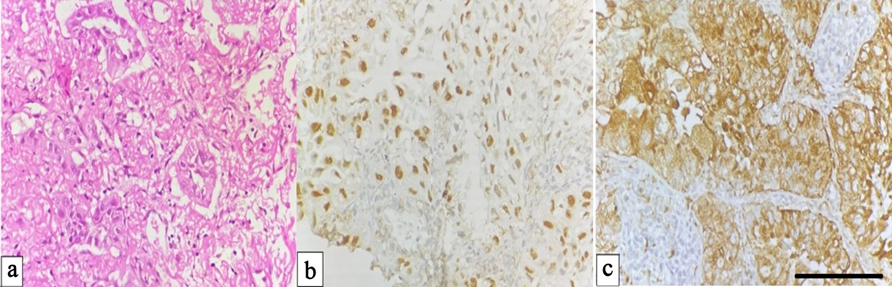
Methods: A prospective study was conducted on 151 lung biopsy specimens over 18 months. All samples were processed using standard histopathological techniques and an IHC panel comprising thyroid transcription factor-1 (TTF-1), napsin A, cytokeratin (CK)7, CK5/6, P63, synaptophysin, and chromogranin. Additional markers (estrogen receptor (ER), progesterone receptor (PR), human epidermal growth factor receptor 2 (HER2), PAX-8, PAX-2, CD10) were applied for metastatic differentiation. Statistical correlation analysis between lineage-specific markers was performed to assess diagnostic concordance.
Results: Adenocarcinoma was the most prevalent subtype (58.3%), followed by neuroendocrine neoplasms (17.2%), metastatic lesions (13.2%), adenosquamous carcinoma (6.0%), and squamous cell carcinoma (2.6%). Rare entities included carcinosarcoma and mucoepidermoid carcinoma (each 1.3%). TTF-1 and napsin A were highly specific for adenocarcinoma (85% and 94%, respectively), whereas CK5/6 and P63 confirmed squamous differentiation (100% each). Synaptophysin and chromogranin were positive in 96% and 100% of neuroendocrine tumors, respectively. Correlation analysis demonstrated strong marker concordance: TTF-1 vs napsin A (r = 0.88, P < 0.001), P63 vs CK5/6 (r = 0.93, P < 0.001), and synaptophysin vs chromogranin (r = 0.87, P < 0.001). All metastatic lesions were TTF-1 negative, confirming their extrapulmonary origin.
Conclusions: Combining IHC with histopathology markedly improves diagnostic precision and consistency in lung tumor classification. Lineage-specific markers validate the IHC panel’s reliability, distinguishing adenocarcinoma, squamous, and neuroendocrine types while differentiating primary from metastatic lesions. This integrative approach supports WHO-aligned precision diagnostics and guides individualized therapeutic strategies.
Published
Issue
Section
License
Copyright (c) 2026 The authors

This work is licensed under a Creative Commons Attribution 4.0 International License.