Elevated E2F6 Expression in Colorectal Cancer Tissues and Its Association With Clinicopathological Features

Authors

  • Da Tong Zeng
  • Ke Jun Wu
  • Jian Di Li
  • Guo Qiang Chen
  • Wei Zhang
  • Zong Yu Li
  • Jing Wen Ling
  • Wei Jian Huang
  • Gang Chen
  • Hui Li

DOI:

https://doi.org/10.14740/wjon2707

Keywords:

Colorectal cancer, E2F transcription factor 6, Clustered regularly interspaced short palindromic repeats, Spatial transcriptomics, Clinicopathological features, Diagnostic biomarker, Tumor budding

Abstract

Background: Colorectal cancer (CRC) is the third most common malignancy worldwide, and the role of E2F transcription factor 6 (E2F6) in CRC remains controversial.

Methods: We analyzed E2F6 mRNA expression across 19 platforms (2,449 CRC patients and 1,328 controls), evaluated expression patterns using single-cell RNA sequencing (scRNA-seq) and spatial transcriptomics, assessed E2F6 dependency using clustered regularly interspaced short palindromic repeats (CRISPR) knockout data from 52 CRC cell lines, and validated protein expression by immunohistochemistry (IHC) in 200 paired CRC and adjacent tissues. Associations between E2F6 and clinicopathological features were analyzed.

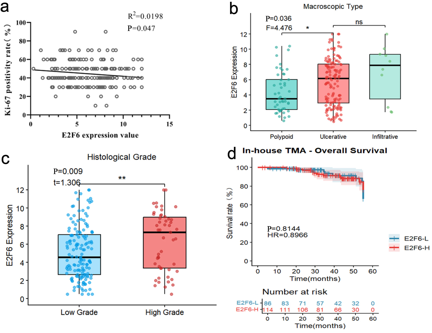
Results: E2F6 was significantly upregulated in CRC versus controls (summary receiver operating characteristic (sROC) area under the curve (AUC) = 0.93), supported by scRNA-seq and spatial transcriptomics. E2F6 knockout suppressed proliferation across CRC cell lines, and IHC confirmed higher E2F6 protein expression (AUC = 0.91). Elevated E2F6 correlated with adverse clinicopathological features including female sex, age ≥ 60 years, advanced T stage, high-grade tumor budding, and higher histological grade.

Conclusions: E2F6 is highly expressed in CRC and is associated with unfavorable clinicopathological features, supporting its potential utility as a diagnostic biomarker and a candidate target for CRC stratification and therapy development.

Author Biographies

  • Da Tong Zeng, Department of Pathology, The First Affiliated Hospital of Guangxi Medical University, Nanning 530021, Guangxi Zhuang Autonomous Region, China

    Department of Pathology, The First Affiliated Hospital of Guangxi Medical University, Nanning 530021, Guangxi Zhuang Autonomous Region, China.

  • Ke Jun Wu, Department of Pathology, The First Affiliated Hospital of Guangxi Medical University, Nanning 530021, Guangxi Zhuang Autonomous Region, China

    Department of Pathology, The First Affiliated Hospital of Guangxi Medical University, Nanning 530021, Guangxi Zhuang Autonomous Region, China.

  • Jian Di Li, Department of Pathology, The First Affiliated Hospital of Guangxi Medical University, Nanning 530021, Guangxi Zhuang Autonomous Region, China

    Department of Pathology, The First Affiliated Hospital of Guangxi Medical University, Nanning 530021, Guangxi Zhuang Autonomous Region, China.

  • Guo Qiang Chen, Department of Pathology, The First Affiliated Hospital of Guangxi Medical University, Nanning 530021, Guangxi Zhuang Autonomous Region, China

    Department of Pathology, The First Affiliated Hospital of Guangxi Medical University, Nanning 530021, Guangxi Zhuang Autonomous Region, China.

  • Wei Zhang, Department of Pathology, The First Affiliated Hospital of Guangxi Medical University, Nanning 530021, Guangxi Zhuang Autonomous Region, China

    Department of Pathology, The First Affiliated Hospital of Guangxi Medical University, Nanning 530021, Guangxi Zhuang Autonomous Region, China.

  • Zong Yu Li, Department of Pathology, The First Affiliated Hospital of Guangxi Medical University, Nanning 530021, Guangxi Zhuang Autonomous Region, China

    Department of Pathology, The First Affiliated Hospital of Guangxi Medical University, Nanning 530021, Guangxi Zhuang Autonomous Region, China.

  • Jing Wen Ling, Department of Pathology, The First Affiliated Hospital of Guangxi Medical University, Nanning 530021, Guangxi Zhuang Autonomous Region, China

    Department of Pathology, The First Affiliated Hospital of Guangxi Medical University, Nanning 530021, Guangxi Zhuang Autonomous Region, China.

  • Wei Jian Huang, Department of Pathology, Redcross Hospital of Yulin City, Yulin 537000, Guangxi Zhuang Autonomous Region, China

    Department of Pathology, Redcross Hospital of Yulin City, Yulin 537000, Guangxi Zhuang Autonomous Region, China

  • Gang Chen, Department of Pathology, The First Affiliated Hospital of Guangxi Medical University, Nanning 530021, Guangxi Zhuang Autonomous Region, China

    Department of Pathology, The First Affiliated Hospital of Guangxi Medical University, Nanning 530021, Guangxi Zhuang Autonomous Region, China.

  • Hui Li, Department of Colorectal and Anal Surgery, The First Affiliated Hospital of Guangxi Medical University, Nanning 530021, Guangxi Zhuang Autonomous Region, China

    Department of Colorectal and Anal Surgery, The First Affiliated Hospital of Guangxi Medical University, Nanning 530021, Guangxi Zhuang Autonomous Region, China

Downloads

Published

2026-05-09

Issue

Section

Original Article

How to Cite

1.
Zeng DT, Wu KJ, Li JD, et al. Elevated E2F6 Expression in Colorectal Cancer Tissues and Its Association With Clinicopathological Features. World J Oncol. 2026;17(3):322-336. doi:10.14740/wjon2707

Similar Articles

31-40 of 104

You may also start an advanced similarity search for this article.