| World Journal of Oncology, ISSN 1920-4531 print, 1920-454X online, Open Access |
| Article copyright, the authors; Journal compilation copyright, World J Oncol and Elmer Press Inc |
| Journal website https://wjon.elmerpub.com |
Original Article
Volume 17, Number 5, October 2026, pages 623-632

Siglec-15 Expression in Gastric Carcinoma and Its Association With Clinicopathological Variables
Sara Al Badra, Abdelkader Battahb, Ahmad Alsughayerc, Fanar Alsmaratd, Khalid Halahlehe, Maher Sughayera, b, f
aDepartment of Pathology and Laboratory Medicine, King Hussein Cancer Center, Amman 11941, Jordan
bSchool of Medicine, University of Jordan, Amman 11941, Jordan
cDepartment of Surgery, King Hussein Cancer Center, Amman 11941, Jordan
dDepartment of Anatomy, Physiology and Biochemistry, Faculty of Medicine, The Hashemite University, Zarqa 13133, Jordan
eDepartment of Medicine, King Hussein Cancer Center, Amman, Jordan
fCorresponding Author: Maher Sughayer, Department of Pathology and Laboratory Medicine, King Hussein Cancer Center, Amman 11941, Jordan
Manuscript submitted February 12, 2026, accepted July 10, 2026, published online September 4, 2026
Short title: Siglec-15 and Clinicopathology in Gastric Carcinoma
doi: https://doi.org/10.14740/wjon2749
| Abstract | ▴Top |
Background: Gastric cancer remains the fifth most common malignancy worldwide and is often diagnosed at advanced stages with limited treatment options. Although immunotherapy targeting the programmed cell death protein-1 (PD-1)/programmed death-ligand 1 (PD-L1) has improved outcomes in several cancers, many patients exhibit resistance, necessitating alternative therapeutic strategies. Sialic acid-binding immunoglobulin-like lectin-15 (Siglec-15), a recently recognized immune suppressor, is overexpressed in tumor cells and tumor-associated macrophages (TAMs), and is largely absent from normal tissues, making it a promising immunotherapy target.
Methods: This tissue microarray (TMA) study retrospectively collected formalin-fixed paraffin-embedded (FFPE) tissue specimens from 2015 to 2019 for 77 gastric cancer patients from King Hussein Cancer Center. Siglec-15 expression patterns in tumor cells and TAMs were analyzed and compared with clinicopathological features and patient outcomes. Survival was assessed using Kaplan–Meier analysis.
Results: Siglec-15 expression was detected in 50.7% of tumor samples and 36.4% of TAMs. Membranous expression was significantly associated with tumor differentiation, with higher expression observed in moderately differentiated tumors (P = 0.048), while cytoplasmic and TAM expression showed no significant associations with clinical variables. Although not statistically significant, positive Siglec-15 expression—both membranous and cytoplasmic—was associated with shorter median overall survival (cytoplasmic: 30.62 vs. 74.92 months, P = 0.1790; membranous: 39.44 vs. 66.07 months, P = 0.5761). Established prognostic indicators such as age, clinical stage, and pT stage were significantly associated with overall survival.
Conclusion: Siglec-15 is frequently expressed in gastric cancer and may contribute to immune evasion within the tumor microenvironment. Although no significant association with prognosis was found, its high expression underscores its potential as a novel immunotherapeutic target. Further studies are warranted to validate these findings and explore Siglec-15 clinical utility.
Keywords: Siglec-15; Gastric carcinoma; Immune checkpoint
| Introduction | ▴Top |
Gastric cancer ranks as the fifth most common cancer globally in both incidence and mortality [1]. The majority of gastric cancer patients are diagnosed at advanced stages, when treatment options are limited and the prognosis is poor [2].
Recent advances in cancer immunotherapy, particularly the blockade of programmed cell death protein-1 (PD-1) and its ligand PD-L1, have revolutionized treatment outcomes for cancer patients by reestablishing anti-tumor immune responses within the tumor microenvironment (TME) [3]. However, many patients exhibit either primary resistance or eventually develop acquired resistance to PD-1/PD-L1 blockades [4]. Moreover, this pathway addresses only a subset of mechanisms underlying tumor-induced immune evasion, underscoring the need for alternative or complementary immunotherapeutic targets [4].
The sialic acid-binding immunoglobulin-like lectin-15 (Siglec-15) is a transmembrane protein primarily expressed on macrophages, dendritic cells, and osteoclasts [5]. Notably, compared to normal tissues, Siglec-15 is markedly overexpressed in cancer cells and tumor-associated macrophages (TAMs) [6]. Within TME, Siglec-15 suppresses cytotoxic T-cell activation and proliferation, aiding immune evasion [7]. It also promotes immune suppression via transforming growth factor-β (TGF-β) secretion through the DAP12–Syk pathway [8]. Siglec-15 and PD-L1 expression have been reported to be mutually exclusive, suggesting that Siglec-15 may serve as a promising immunotherapeutic target in PD-L1-negative tumors or in patients who are resistant to PD-1/PD-L1 blockade [8].
Siglec-15 expression has been observed in several cancers, including breast cancer, pancreatic adenocarcinoma, head and neck squamous cell carcinoma, and uterine carcinosarcoma [9]. However, current data on its expression frequency in gastric cancer, its prognostic value, and its association with clinicopathological features are limited and controversial [10]. Therefore, this study aimed to investigate Siglec-15 expression in gastric cancer and examine its association with clinicopathological features and patient prognosis.
| Materials and Methods | ▴Top |
Patients and samples
Following approval from the Institutional Review Board (IRB), formalin-fixed paraffin-embedded (FFPE) tissue blocks were obtained from patients who underwent partial or total gastrectomy at King Hussein Cancer Center (KHCC) between 2010 and 2018. A total of 100 cases of 125 available cases were randomly selected for initial review. For each selected case, corresponding pathology reports, FFPE tumor blocks, and hematoxylin and eosin (H&E)-stained sections were thoroughly examined. Additional clinical data, including overall survival, clinical stage, and smoking history, were extracted from patients’ medical records.
Upon preliminary review, patients diagnosed with non-gastric cancers or with gastric tumors other than adenocarcinomas were removed (n = 8). To preserve the integrity of tissue samples during tissue microarray (TMA) construction, blocks with significant fragmentation were also excluded (n = 7). The remaining 85 cases were examined by one of the researchers (MAS) using several H&E slides. The precise tissue region of interest was determined, and the block with the greatest cancer cell count was selected for TMA production after the examination. Cases with a low number of cancer cells on all available slides were also eliminated to decrease the risk of false-negative results (n = 8). The remaining 77 cases were used to make the TMA blocks. All relevant clinicopathological and histological data were documented in an organized Excel spreadsheet for subsequent analyses.
TMA
Four TMA blocks were constructed and utilized in this study, each containing between 20 and 30 tissue cores, along with at least one control core derived from tonsillar tissue. From each TMA block, two 4 µm sections were obtained. The first section was stained with H&E to assess the adequacy and integrity of the tumor cores. The second section, intended for immunohistochemistry (IHC), was made on positively charged slides.
Duplicate cores from the same case were included in instances where tissue quality was suboptimal or the tumor location within the core was uncertain. If a core was lost during sectioning, a replacement core from the corresponding original FFPE block was extracted and incorporated into the final TMA, ensuring near-complete representation of the initially selected cases.
IHC procedure and evaluation
According to the manufacturer’s recommendations, TMA slides were immunostained using an anti-Siglec-15 antibody (rabbit polyclonal antibody PA5-116878, Thermo Fisher Scientific, USA) on the Ventana Benchmark Ultra platform (Roche, Tucson, AZ). Both positive and negative controls were included on every slide to ensure staining reliability.
The IHC evaluation was performed by one of the researchers (MAS) using a manual precise count method by examining the cores at × 200 magnification and counting no fewer than 100 cancer cells and TAM cells in each case using a light microscope. Siglec-15-positive immunolabeling was predominantly observed as membranous and/or cytoplasmic staining in tumor cells, while TAM exhibited linear membranous, diffuse, and/or punctate cytoplasmic staining. The cut-off values used in the study are as follows: 40% for cytoplasmic staining, 20% for membranous staining, and 20% for TAM staining. A positive cell for TAM and cytoplasmic staining includes any intensity and for membranous staining any intensity and either partial or complete membranous staining.
Statistical analyses
Descriptive statistics were employed to summarize the patients’ demographic and clinical characteristics. Comparisons between Siglec-15 positive and negative cases with respect to various risk factors were conducted using Fisher’s exact test. For the survival analysis, Kaplan–Meier curves were generated, with a significance level set at P ≤ 0.05. All statistical analyses were performed using SAS version 9.4 (SAS Institute Inc., Cary, NC).
IRB approval
The IRB at KHCC has approved this study under the Reference No. 24 KHCC 130. The study was conducted in compliance with the ethical standards of the responsible institution on human subjects as well as with the Helsinki Declaration.
| Results | ▴Top |
Patient characteristics and expression of Siglec-15
The study included predominantly male patients (n = 48, 62.3%). The median age for the study cohort was 65 years and the interquartile was 25. At the time of analysis, 44 patients (57.1%) were deceased. According to Laurén’s classification, the diffuse and indeterminate subtypes were the most prevalent (n = 58, 75.3%), while the intestinal subtype accounted for 19 cases (24.7%). The majority of tumors (54.5%) were poorly differentiated, while 42.9% were moderately differentiated. Among 61 patients with documented clinical stages, 40 cases (65.6%) were diagnosed at stage III or higher. Gastritis was present in 13 cases (16.9%) (Table 1).
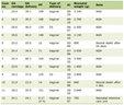 Click to view | Table 1. Demographic, Clinicopathological, and Molecular Characteristics of Gastric Cancer Patients |
Siglec-15 expression was detected in the cytoplasm and/or membranes of gastric cancer cells in 39 patients (50.65%), including 14 females (18.18%) and 25 males (32.47%). Specifically, Siglec-15 was observed in the membranes of tumor cells in 26 patients (33.8%) and in the cytoplasm of 33 patients (42.9%). Additionally, Siglec-15 expression was detected in TAMs in 28 patients (36.4%) (Table 1, Fig. 1).
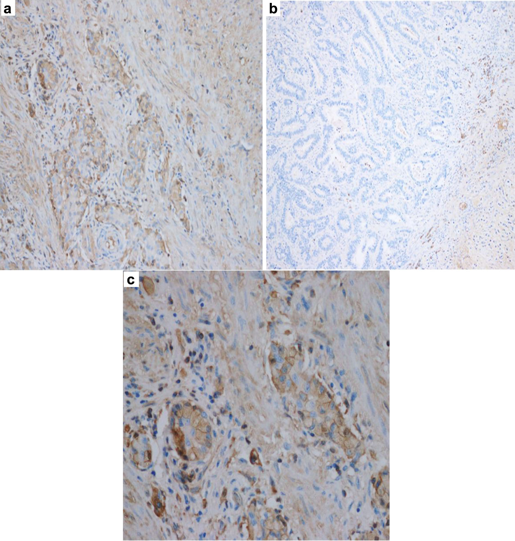 Click for large image | Figure 1. Representative immunohistochemistry images of Siglec-15 expression in gastric cancer tissues. (a) Positive Siglec-15 expression. (b) Negative Siglec-15 expression. (c) Positive membranous and cytoplasmic expression in tumor cells and Positive expression in tumor-associated macrophages (TAMs). |
Association of Siglec-15 expression with clinicopathological factors in gastric cancer
Membranous Siglec-15 expression in gastric cancer cells was significantly associated with tumor differentiation (P = 0.048), with higher expression observed in moderately differentiated tumors (Table 2). However, no significant associations were found with age, gender, clinical stage, signet ring cell histology, tumor location, neoadjuvant therapy, gastritis, H. pylori infection, lymphovascular invasion, PD-L1 status, pT, pN, pM, or Laurén classification. Cytoplasmic Siglec-15 expression did not demonstrate significant associations with clinicopathological factors, though a trend was observed toward higher expression in tumors with lymphovascular invasion (P = 0.075). Similarly, Siglec-15 expression in TAMs showed no significant association with any clinicopathological factors (Table 3).
Click to view | Table 2. Membranous and Cytoplasmic Siglec-15 Expression and Its Association With Various Clinical Variables in Gastric Cancer Patients |
Click to view | Table 3. TAM Siglec-15 Expression and Its Association With Various Clinical Variables in Gastric Cancer Patients |
Siglec-15 expression and overall survival
Patients with positive cytoplasmic or membranous Siglec-15 expression had shorter median overall survival, though the differences were not statistically significant (P = 0.1790 and P = 0.5761, Fig. 2). Median overall survival was 30.62 months for patients with positive cytoplasmic Siglec-15 expression compared to 74.92 months for patients with negative cytoplasmic Siglec-15 expression, while patients with positive Siglec-15 membranous expression had a median survival of 39.44 months compared to 66.07 months for patients with negative membranous Siglec-15 expression.
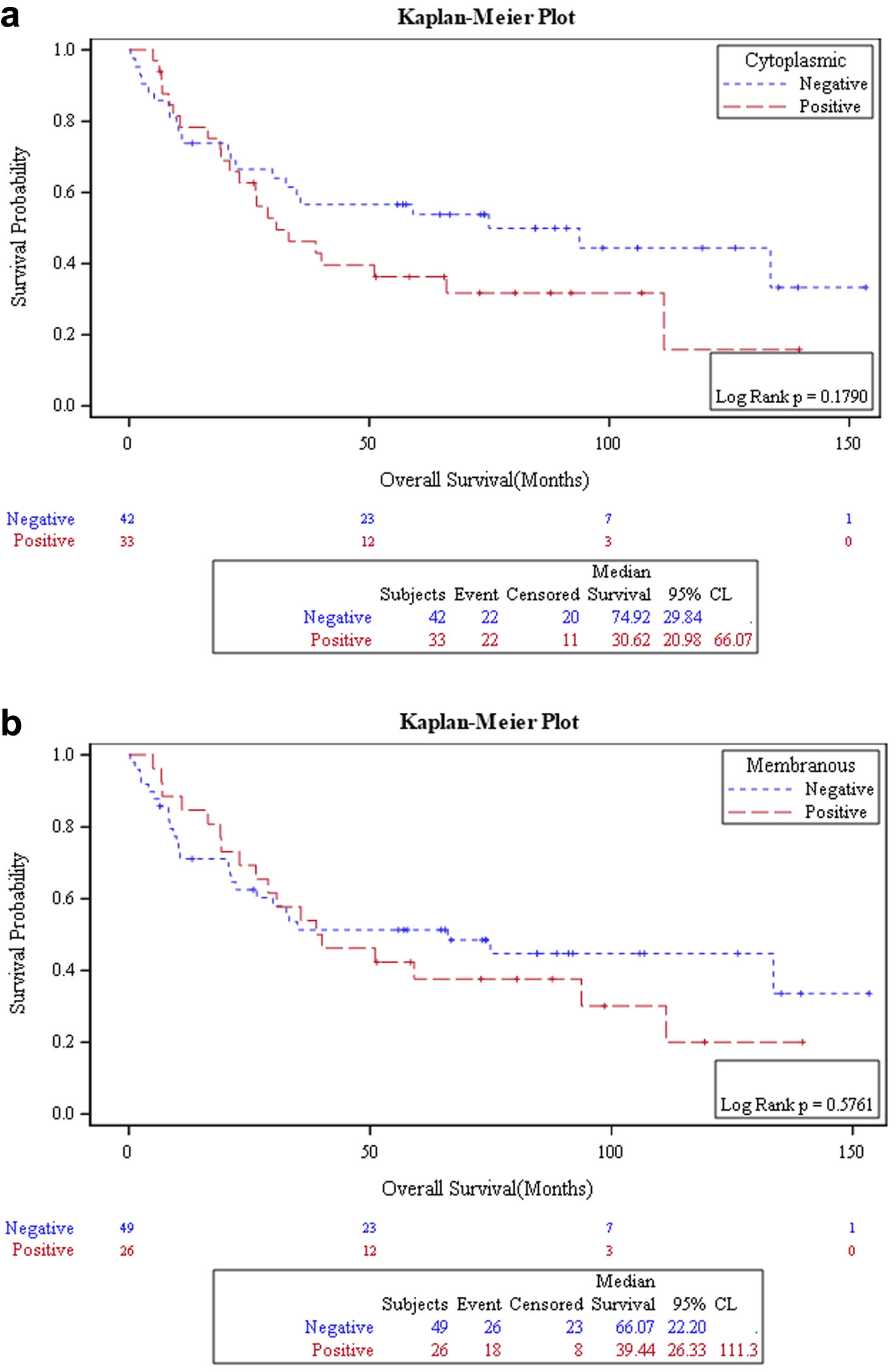 Click for large image | Figure 2. Kaplan–Meier curve analysis of overall survival in gastric cancer patients. (a) Association between cytoplasmic Siglec-15 expression and overall survival. (b) Association between membranous Siglec-15 expression and overall survival. |
The 5-year survival rate was lower in patients with positive cytoplasmic Siglec-15 expression (36.3%, 95% confidence interval (CI): 20.5–53.7) compared to patients with negative cytoplasmic Siglec-15 expression (53.8%, 95% CI: 38.5–68.7). A similar trend was observed for membranous Siglec-15 expression, with 5-year survival rates of 37.6% (95% CI: 20.0–57.0) in positive membranous Siglec-15 expression patients versus 51.3% (95% CI: 37.1–65.4) in negative membranous Siglec-15 expression patients.
Several established clinicopathological factors were significantly associated with overall survival in gastric cancer patients. Advanced age was found to be significantly associated with poorer median overall survival (P = 0.0108, Fig. 3a). Additionally, patients diagnosed at early stages (I/II) experienced significantly longer median overall survival compared to patients diagnosed at advanced stages (III/IV) (P = 0.0049, Fig. 3b). Furthermore, a higher pT stage was significantly associated with decreased median overall survival (P = 0.0252, Fig. 3c).
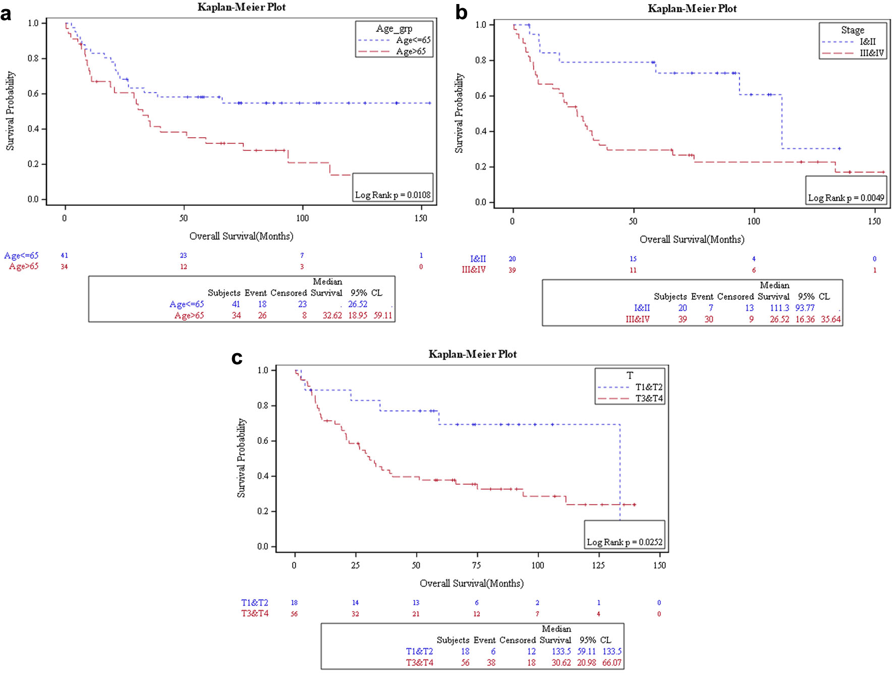 Click for large image | Figure 3. Kaplan–Meier curve analysis of overall survival in gastric cancer patients. (a) Association between age and overall survival. (b) Association between clinical stage and overall survival. (c) Association between pathological T stage and overall survival. |
| Discussion | ▴Top |
Siglec-15 is a promising novel target for immunotherapy to restore anti-tumor immunity within the TME. Unlike other immune suppressors, Siglec-15 is poorly expressed in normal tissues but highly expressed in TAMs and cancer cells, making it a selective target for therapy [7, 11, 12]. Preclinical studies in mouse models have demonstrated that Siglec-15 inhibition enhances anti-tumor immune responses within the TME [7]. In our cohort of gastric cancer patients, cytoplasmic and/or membranous Siglec-15 expression was observed in 50.7% of tumor samples, and 36.4% of cases exhibited Siglec-15 positivity in TAMs. To our knowledge, this is the first study to evaluate Siglec-15 expression in gastric cancer within the Jordanian population. Our results are consistent with those of Quirino et al [10], who reported Siglec-15 expression in 61.9% of gastric cancer tumors and 42.5% of TAMs. Our results are further supported by transcriptomic data indicating elevated Siglec-15 mRNA levels in gastric cancer [9].
The overexpression of Siglec-15 has been identified in a range of malignancies, including bladder, breast, thyroid, esophageal, renal, hepatocellular, and lung cancers [6, 9]. For instance, Yang et al [13] found Siglec-15 expression on both tumor cells and TAMs in clear cell renal cell carcinoma. Additionally, Siglec-15 expression was present in both TAMs (13.3%) and neoplastic cells (22.8%) of lung cancer samples [7]. Similarly, Li et al [9] observed membranous Siglec-15 expression in 16.5% of lung adenocarcinoma cases. Shafi et al [11] further demonstrated significant Siglec-15 expression in TAMs and tumor cells across multiple cancers, including breast, lung, head and neck, and bladder cancers. Notably, TAM expression consistently exceeded tumor cell expression in those cancers [11]. In contrast, our findings revealed a higher frequency of Siglec-15 positivity in cancer cells than in TAMs. This discrepancy may stem from methodological differences, including the use of varying antibody types and different cut-off values.
Siglec-15 exerts immunosuppressive effects through mechanisms distinct from PD-1/PD-L1, offering potential therapeutic advantages for patients who do not respond to anti-PD-1/PD-L1 therapies or exhibit resistance [7]. Siglec-15 and PD-L1 typically showed mutually exclusive expression [7, 11], although Yang et al [13] found no association in clear cell renal cell carcinoma. In our gastric cancer cohort, no significant association was observed between Siglec-15 and PD-L1, likely due to the small sample size.
In this study, a trend toward poorer prognosis with positive Siglec-15 expression was observed, although the association did not reach statistical significance. Notably, 5-year survival rates were markedly lower in patients with cytoplasmic (36.3%) or membranous (37.6%) Siglec-15 expression, compared to the survival rate in patients with negative Siglec-15 expression (53.8% and 51.3%, respectively). Li et al [9] reported similar findings, linking Siglec-15 expression to unfavorable outcomes. In contrast, studies by Quirino et al [10] in gastric cancer and Hao et al [14] in lung cancer found a positive association between positive Siglec-15 expression and patient survival. These discrepancies underscore the need for further large-scale, multi-center investigations to clarify the prognostic significance of Siglec-15 across tumors.
Siglec-15 expression was not significantly associated with most clinicopathological features in our cohort. The only significant finding was an association between membranous Siglec-15 expression and tumor differentiation (P = 0.048), with higher frequency of positivity observed in moderately differentiated tumors. This is consistent with studies by Quirino et al, Shafi et al, and Yang et al [10, 11, 13]. While previous reports have linked Siglec-15 to factors such as lymphovascular invasion and TNM stage, findings remain inconsistent [10, 11, 15]. In our data, cytoplasmic expression showed a non-significant trend toward association with lymphovascular invasion (P = 0.075) and absence of metastasis (P = 0.068).
This study investigated Siglec-15 expression in gastric cancer patients to assess its potential as an immunotherapeutic target and to explore its association with clinicopathological features. Limitations of this study include the small sample size, reliance on a single method for detecting Siglec-15 expression, and the use of anti-Siglec-15 polyclonal antibodies without independent validation against an alternative detection method. Finally, the cohort was drawn from a single ethnic population, which may limit the generalizability of these findings to other ethnicities, particularly Asian populations in whom gastric cancer prevalence is considerably higher.
In conclusion, this study found elevated Siglec-15 protein expression in half of the patients, with a significant association only with histological grade. While Siglec-15 expression in cancer cells was linked to lower overall survival, this relationship was not statistically significant. Survival analysis showed lower survival rates in patients with older age (P = 0.0108), higher clinical stage (P = 0.0049), and higher pT stage (P = 0.0252). Additionally, cytoplasmic Siglec-15 expression showed trends toward associations with lymphovascular invasion (P = 0.075) and absence of metastasis (P = 0.068). Future studies with larger sample sizes are needed to better evaluate the relationship between Siglec-15 expression and clinicopathological features.
Acknowledgments
None to declare.
Financial Disclosure
This research did not receive any specific grant from any funding agencies.
Conflict of Interest
The authors declared no potential conflicts of interest with respect to the research, authorship, and/or publication of this article.
Informed Consent
Informed consent was waived by the IRB.
Author Contributions
Conceptualization: MS and AB; data collection: SA, AA, and KH; writing—original draft: SA; writing—review and editing: all authors; supervision: MS.
Data Availability
The data supporting the findings of this study are available from the corresponding author upon reasonable request.
| References | ▴Top |
- Bray F, Laversanne M, Sung H, Ferlay J, Siegel RL, Soerjomataram I, Jemal A. Global cancer statistics 2022: GLOBOCAN estimates of incidence and mortality worldwide for 36 cancers in 185 countries. CA Cancer J Clin. 2024;74(3):229-263.
doi pubmed - Pasechnikov V, Chukov S, Fedorov E, Kikuste I, Leja M. Gastric cancer: prevention, screening and early diagnosis. World J Gastroenterol. 2014;20(38):13842-13862.
doi pubmed - Jia L, Zhang Q, Zhang R. PD-1/PD-L1 pathway blockade works as an effective and practical therapy for cancer immunotherapy. Cancer Biol Med. 2018;15(2):116-123.
doi pubmed - Ribas A, Wolchok JD. Cancer immunotherapy using checkpoint blockade. Science. 2018;359(6382):1350-1355.
doi pubmed - Angata T, Tabuchi Y, Nakamura K, Nakamura M. Siglec-15: an immune system Siglec conserved throughout vertebrate evolution. Glycobiology. 2007;17(8):838-846.
doi pubmed - Li QT, Huang ZZ, Chen YB, Yao HY, Ke ZH, He XX, Qiu MJ, et al. Integrative analysis of Siglec-15 mRNA in human cancers based on data mining. J Cancer. 2020;11(9):2453-2464.
doi pubmed - Wang J, Sun J, Liu LN, Flies DB, Nie X, Toki M, Zhang J, et al. Siglec-15 as an immune suppressor and potential target for normalization cancer immunotherapy. Nat Med. 2019;25(4):656-666.
doi pubmed - Takamiya R, Ohtsubo K, Takamatsu S, Taniguchi N, Angata T. The interaction between Siglec-15 and tumor-associated sialyl-Tn antigen enhances TGF-beta secretion from monocytes/macrophages through the DAP12-Syk pathway. Glycobiology. 2013;23(2):178-187.
doi pubmed - Li B, Zhang B, Wang X, Zeng Z, Huang Z, Zhang L, Wei F, et al. Expression signature, prognosis value, and immune characteristics of Siglec-15 identified by pan-cancer analysis. Oncoimmunology. 2020;9(1):1807291.
doi pubmed - Quirino MWL, Pereira MC, Deodato de Souza MF, Pitta IDR, Da Silva Filho AF, Albuquerque MSS, Albuquerque APB, et al. Immunopositivity for Siglec-15 in gastric cancer and its association with clinical and pathological parameters. Eur J Histochem. 2021;65(1):3174.
doi pubmed - Shafi S, Aung TN, Xirou V, Gavrielatou N, Vathiotis IA, Fernandez A, Moutafi M, et al. Quantitative assessment of Siglec-15 expression in lung, breast, head, and neck squamous cell carcinoma and bladder cancer. Lab Invest. 2022;102(10):1143-1149.
doi pubmed - Sun J, Lu Q, Sanmamed MF, Wang J. Siglec-15 as an emerging target for next-generation cancer immunotherapy. Clin Cancer Res. 2021;27(3):680-688.
doi pubmed - Yang WB, Qin CP, Du YQ, Han SC, Xu T. Siglec-15 promotes progression of clear renal cell carcinoma. Chin Med J (Engl). 2021;134(21):2635-2637.
doi pubmed - Hao JQ, Nong JY, Zhao D, Li HY, Su D, Zhou LJ, Dong YJ, et al. The significance of Siglec-15 expression in resectable non-small cell lung cancer. Neoplasma. 2020;67(6):1214-1222.
doi pubmed - Chen G, Luo S, Kozlovich S, Lazarus P. Association between glucuronidation genotypes and urinary NNAL metabolic phenotypes in smokers. Cancer Epidemiol Biomarkers Prev. 2016;25(7):1175-1184.
doi pubmed
This article is distributed under the terms of the Creative Commons Attribution 4.0 International License (CC BY 4.0), which permits unrestricted use, distribution, and reproduction in any medium, including commercial use, provided the original work is properly cited.
World Journal of Oncology is published by Elmer Press Inc.