| World Journal of Oncology, ISSN 1920-4531 print, 1920-454X online, Open Access |
| Article copyright, the authors; Journal compilation copyright, World J Oncol and Elmer Press Inc |
| Journal website https://wjon.elmerpub.com |
Original Article
Volume 17, Number 6, December 2026, pages 912-922

Relationship Between Estrogen-Induced BRCA2 and Development of Gastric Cancer
Takuma Hayashia, c, Ikuo Konishia, b
aCancer Medicine, National Hospital Organization, Kyoto Medical Center, Kyoto City, Kyoto 612-8555, Japan
bKyoto University School of Medicine, Kyoto City, Kyoto 606-8501, Japan
cCorresponding Author: Takuma Hayashi, Cancer Medicine, National Hospital Organization, Kyoto Medical Center, Kyoto City, Kyoto 612-8555, Japan
Manuscript submitted September 9, 2026, accepted October 23, 2026, published online November 11, 2026
Short title: BRCA2 Expression Influences Development of GC
doi: https://doi.org/10.14740/wjon2875
| Abstract | ▴Top |
Background: Gastric cancer (GC) is the fourth most common malignancy, affecting one in 11 men (9.1%) and one in 23 women (4.38%). Helicobacter pylori (H. pylori) infection and smoking are the primary risk factors for GC. Individuals who have multiple other family members with GC are also at higher risk owing to lifestyle and genetic factors. H. pylori-eradicating medications can reduce the risk of disease. The incidence of GC increases after 50 years of age, particularly among men; however, the reason for the differing incidence rates between the sexes remains unclear.
Methods: To clarify the reasons for sex differences in GC incidence rates from a molecular pathological perspective, we utilized cancer genome profiling (CGP) and information on BRCA2 expression levels obtained by immunohistochemistry and the number of GC cases in the family.
Results: We investigated the incidence of GC in families with hereditary breast and ovarian cancer (HBOC) and found it to be 4.2-fold higher than that in non-HBOC families. In families with versus without HBOC, men accounted for 74.57% and 53.67% of all GC cases, respectively, indicating that men are more likely to develop GC in the former group. Furthermore, estrogen stimulation induced breast cancer type 2 (BRCA2) expression in genetically modified mice as well as in cultured cells, and low-grade atypical cells were observed in the gastric mucosal tissues of female mice that underwent ovarian resection. Additional research experiment revealed that stimulation by the female hormone estrogen induces the expression of BRCA2, which suppresses the development of GC. Clinical studies have also shown that patients with GC who have high BRCA2 expression exhibit longer survival rates than those in whom BRCA2 expression is low.
Conclusions: Overall, the data suggest that the higher mortality rate of GC in men than in women may be due to a protective effect of BRCA2.
Keywords: Gastric cancer; BRCA 1/2; Helicobacter pylori; Pathogenic variant
| Introduction | ▴Top |
Gastric cancer (GC) involves uncontrolled growth of cancerous mucosal cells in the lining of the stomach and can take several years to become detectable. Helicobacter pylori (H. pylori) is a gastric bacterial pathogen associated with human GC; Sugano et al reported that patients with H. pylori-associated gastritis and gastric ulcers have a higher risk of GC than those with duodenal ulcers, indicating that gastric atrophy remains a risk factor even after H. pylori eradication therapy. Clinical research conducted by a Japanese medical team concluded that careful monitoring, such as endoscopic examination, is required after the successful eradication of H. pylori in patients who are considered to be at a higher risk [1].
H. pylori is a spiral-shaped bacterium that inhabits the gastric lining. Even in a highly acidic gastric environment, it survives by using the enzyme urease to break down urea into the more neutral ammonia. There is a strong association between H. pylori and stomach cancer; H. pylori is among the main causes of GC. The cytotoxin-associated gene A (CagA) protein of H. pylori, to which gastric epithelial cells are exposed following bacterial type IV secretion, is an oncoprotein that induces malignant neoplasms in mammals. Genetic alterations of H. pylori, most commonly in CagA and vacuolating cytotoxin, also occur; CagA-positive H. pylori strains increase the risk of atrophic gastritis and GC [2]. Numerous clinical studies are being conducted to identify other genes associated with GC development.
Breast cancer susceptibility gene 1 (BRCA1) is a tumor suppressor gene located on human chromosome 17 and is involved in DNA repair and cell cycle regulation. BRCA1 encodes a protein that helps repair damaged DNA, thereby maintaining the stability of the genetic information in the cell. If an abnormality occurs during cell division, BRCA1 either suppresses cell proliferation or induces apoptosis. The association between BRCA1 mutations and a higher risk of cancer development has previously been established; a germinal mutation in the BRCA1 gene significantly increases the risk of developing hereditary breast and ovarian cancer (HBOC) owing to reduced DNA repair function.
Approximately 50% of the global population is infected with H. pylori, and chronic infection of the gastric mucosa is closely associated with the development of atrophic gastritis and gastric ulcers. In developed countries, H. pylori infection increases the risk of GC at least sixfold. H. pylori strains that are CagA-positive cause more severe atrophic gastritis than CagA-negative counterparts and are more strongly associated with the development of GC. H. pylori infection has also been suggested to be involved in the development of gastric mucosa-associated lymphoid tissue lymphoma [3].
Momozawa et al used next-generation sequencing to compare the genes of patients with GC to those of healthy individuals and to identify genetic biomarkers for this disease. They reported significantly more pathogenic variants (PVs) in germline BRCA1 (gBRCA1) and/or gBRCA2 in patients with GC than in healthy individuals. However, in the four cohorts of patients without CagA derived from H. pylori including gBRCA1/2 wild-type (wt), gBRCA1/2 PVs, gBRCA1 PVs/gBRCA2 wt, and gBRCA1 wt/gBRCA2 PVs, the incidence of GC affliction by 80 years of age was < 10%. Moreover, the incidence of GC among individuals without gBRCA1/2 PVs who were infected with CagA-positive H. pylori was low. These clinical data suggested that the incidence of GC is high among individuals with BRCA1/2 PVs. A large-scale clinical study by Momozawa et al showed that patients with gBRCA1/2 PVs infected with H. pylori had a ≥ 40% incidence rate of GC by 80 years of age. However, healthy individuals with gBRCA1/2 wt had a 10% incidence of GC by 80 years of age despite H. pylori infection status [4, 5].
| Materials and Methods | ▴Top |
Producing GantgBrca1 and GantgBrca2 mice
GantgBrca1 and GantgBrca2 mice were created by crossing C57BL/6JtgBrca1 or C57BL/6JtgBrca2 mice with the Gan mice (kindly provided by Dr. Oshima M (Kanazawa University School of Medicine, Kanazawa, Ishikawa, Japan)). The Gan mice are compound transgenic mice created by crossing C2mE mouse and Wnt mouse. In the Gan mice, both the COX-2/PGE2 pathway and Wnt signaling are activated in the gastric mucosa. The Gan mice spontaneously develop ductal GC accompanied by an inflammatory response, with 100% efficiency. The Gan mice are considered as models of human GC, wherein tumors develop through the interaction of Wnt signal activation and inflammatory responses.
C57BL/6JtgBrca1 and C57BL/6JtgBrca2 mice used to obtain GantgBrca1 and GantgBrca2 mice were produced as follows.
MMTV-BRCA1 transgenic constructs
Diagram of the BRCA1 cDNAs used for the generation of transgenic animals is shown in Section of “Generation and Maintenance of BRCA2 Transgenic Mice” (Supplementary Material 1, wjon.elmerpub.com). In MMTV-BRCA1 transgenic constructs, the expression of wild-type BRCA1 is controlled by the mouse mammary tumor virus (MMTV)-long terminal repeat (LTR) promoter. The BRCA1 cDNAs were each inserted into the third exon of the rabbit β-globin gene (β-g). The bar indicates the Really Interesting New Gene finger motif, the hatched region corresponds to the nuclear localization signals, and the negative symbols (−) indicate the negatively charged C-terminal domain with transactivation function.
Generation and maintenance of BRCA1 transgenic (C57BL/6JtgBrca1) mice
The Xho I BRCA1 fragments were microinjected into the male pronucleus of C57BL/6x DBA/2 F1 fertilized mice embryos (obtained from CLEA Japan, Inc., Tokyo, Japan) and implanted into pseudo pregnant ICR surrogate mice at the Transgenic/Embryonic Stem Cell Shared Resource Facility (Japan) to obtain founder mice (Supplementary Material 2, wjon.elmerpub.com). Founder mice were bred with C57BL/6J mice (obtained from CLEA Japan, Inc. Meguro, Tokyo, Japan) to establish the C57BL/6JtgBrca1 transgenic mice line.
CMV-BRCA2 transgenic constructs
To construct p236BRCA2, the pcDNA3 vector was first modified by inserting a 236-bp fragment of the 5' untranslated region of BRCA2 between the KpnI and NotI sites. The assembled full-length BRCA2 cDNA was then inserted at the XhoI site of this plasmid. The 5' UTR of BRCA2 was obtained by RT-PCR using primers 5'-GGTACCGGTG GCGCGAGCTT CTGA-3' and 5'-GCGGCCGCAACTACGATATTCCTCCAAT-3'. The pcDNA3 236HSC WT (BRCA2) was a gift from Mien-Chie Hung (Addgene plasmid #16246; RRID:Addgene_16246) [6] (Supplementary Material 2, wjon.elmerpub.com).
Generation and maintenance of BRCA2 transgenic (C57BL/6JtgBrca2) mice
The BRCA2 gene was generated using recombineering, and transgenic mice were generated as described previously in the section describing BRCA1 transgenic mice.
All animals used in these studies were handled in strict compliance with Shinshu University School of Medicine, Animal Care Committee regulations (Approved Number: 567-5).
Cancer gene panel profiling
The study included 183 families with HBOC encompassing 4,847 participants, among whom 173 had GC, as well as 189 families without HBOC encompassing 4,755 participants, among whom 41 had GC. Between December 2019 and August 2025, 47,211 novel treatments were investigated using cancer genome panel testing (FoundationOne CDx test, Foundation Medicine, Inc., Cambridge, MA, USA, n = 38,981 and OncoGuide™ NCC Oncopanel test, Riken Genesis, Yokohama, Kanagawa, Japan, n = 8,230) at Japanese national universities. We examined a novel treatment method using cancer genome panel testing in 2,489 Japanese patients with advanced-stage or metastatic GC. The clinical research project was approved by the institutional review board (IRB; Approval Number R34005, H31-cancer-2).
Diagnostic testing for H. pylori infection
We used highly sensitive urea breath and stool H. pylori antigen tests combined with the ABC screening method. We stained tissue sections obtained during surgery with hematoxylin and eosin or Giemsa, and observed them under a microscope. Direct observation allows for the detection of H. pylori even if the bacterium is in the coccoid form (spherical bacteria) that cannot be cultured and has no urease activity.
Western blotting
The expression of estrogen receptor α and BRCA2 in gastric adenocarcinoma cells (AGS cells and MKN28 cells), cultured breast cancer (MCF7), and cervical cancer (HeLa) cells were confirmed using SDS-PAGE and western blot analyses. Two types of gastric adenocarcinoma cells (AGS cells and MKN28 cells) (105 cells each) were seeded in a 10-cm chalet and cultured at 37 °C for 12 h. After 12 h of culture, four different concentrations of estrogen (Cat. #056-04044, FUJIFILM Wako Chemicals, Osaka City, Osaka, Japan) and progesterone (Cat. #160-24511, FUJIFILM Wako Chemicals, Osaka City, Osaka, Japan) were added to the culture medium to achieve final concentrations of 0, 5, 20, and 50 nM, and cultured for another 12 h. Subsequently, each cultured cell was treated with a lysate solution (Cat. #R0278-50ML, Merck KGaA, Darmstadt, Germany). To examine the estrogen receptor α and BRCA2 expression status in each cultured cell in response to estrogen and progesterone stimulation, analysis was performed using a standard western blot protocol.
The cells were maintained at 37 °C in a humidified incubator with 5% CO2. For the hypoxic culture, the cells were cultured in a low oxygen incubator with a gas mixture containing 1% O2, 5% CO2, and 94% N2. We purchased MKN28 cells (JCRB0253 MKN28) from National Institutes of Biomedical Innovation, Health and Nutrition (Kawasaki, Kanagawa, Japan). We purchased AGS cells (CRL-1739), MCF7 cells (HTB-22), and HeLa cells (CRM-CCL-2) from ATCC/American Type Culture Collection (Manassas, Virginia, USA).
Reverse transcription-polymerase chain reaction analysis (RT-PCR)
The expressions of BRCA2, estrogen receptor α, and β-actin transcripts were examined using RT-PCR. Total RNA was prepared from human gastric adenocarcinoma cells (AGS cells and MKN28 cells), cultured breast cancer (MCF7), and cervical cancer (HeLa) cells using TRIzol reagent according to the manufacturer’s protocol (Invitrogen Co., CA, USA). The RNA was reverse-transcribed with the Superscript II enzyme (Invitrogen Co.) and single stranded cDNA was used for amplification. BRCA2, estrogen receptor α, and β-actin transcripts were subjected to PCR with the appropriate primer sets following a program of 35 cycles at 94 °C for 30 s, 60 °C for 30 s, and 72 °C for 1.5 min with an additional 5 min for the extension of transcripts. The primers were used as follows: human BRCA2 (F 5′-GGCTTCAAAAAGCACTCCAGATG-3′, R 5′-GGATTCTGTATCTCTTGACGTTCC-3′), human estrogen receptor α (F 5′-CCACTATGGTGTGGCATCCTGT-3′, R 5′-GGTGATCTCACACTCGTTGGAG-3′), and β-actin (F 5′-CACCATTGGCAATGAGCGGTTC-3′, R 5′-AGGTCTTTGCGGATGTCCACGT-3′). These experiments were conducted at Shinshu University in accordance with local guidelines (Approval No. 4737, No. 150, and No. M192).
Immunostaining
We investigated the expression status of estrogen receptor α and BRCA2 in the mucosal layer of male and female normal stomach tissue. Each gastric mucosal tissue sample was fixed with buffered formalin for 12 h. Afterward, each tissue sample was embedded in a plastic box with paraffin. Each paraffin-embedded tissue was microtome-assisted to create 5 µm thick sections, and then thin section slides were prepared. The thin-sectioned slides were saved in the cryoprotection solution. Slices were transferred from the cryoprotection solution to 0.1 M phosphate-buffered saline (PBS) at room temperature, and then rinsed three times for 10 min each, with 0.1 M PBS. Slices were incubated for 30 min in endogenous peroxidase blocking solution to inactivate endogenous peroxidase, and then rinsed three times, 10 min each, with 0.1 M PBS. Optionally, antigen retrieval was performed. Slices were incubated for 20 min with 2 N HCl at 37 °C, and rinsed in 0.1 M borate buffer (8.5 pH) for 10 min, three times for 10 min each, with ice-cold 0.1 M PBS. Slices were incubated for 2 h at room temperature with PBS++ (blocking solution), and incubated with anti-estrogen receptor α ppbsrimary antibody (mouse host) (Cat. #12255-1-AP, Proteintech Group, Inc., Rosemont, IL, USA) and anti-BRCA2 primary antibody (mouse host) (Cat. #29450-1-AP, Proteintech Group, Inc., Rosemont, IL 60018, USA) at a concentration of 1:250 in PBS+ overnight at 4 °C. On day 2, the slices were rinsed three times for 10 min each with 0.1 M PBS, incubated with 1:250 HRP-conjugated secondary antibody (anti-mouse) (Cat. #G-21234, Invitrogen, Thermo Fisher Scientific, Waltham, MA USA) in PBS+ for 2–4 h at room temperature, rinsed three times for 10 min each with 0.1 M PBS, transferred to DAB peroxidase (HRP) substrate solution, and incubated for 2–10 min. When slices became dark grey, the tissue was visualized with a microscope. If positive cells are present, they were rinsed three times (for 15 min each) with tap water (to reduce background), washed three times for 10 min each with 0.1 M PBS, carefully mounted on gelatinized slides using a soft brush, air dried overnight at room temperature, counterstained with permanent mounting medium added and coverslips placed, and stored at 4 °C for up to 6 months.
Measurement of estrogen levels
For a blood test, a health care professional will take a blood sample from a vein in your arm, using a small needle. After the needle is inserted, a small amount of blood will be collected into a test tube or vial. Asuka Pharmaceutical Medical Co., Ltd. (Fujisawa-city, Kanagawa, Japan) has developed numerous steroid measurement methods using LC-MS/MS. In particular, our methods feature high-sensitivity measurement achieved through purification operations that remove impurities that reduce detection sensitivity in derivatization and mass spectrometry. This technology has enabled ultra-high-sensitivity measurement of estradiol (E2) and estrone (E1).
Statistical analyses
The differences between low-BRCA2 (235 cases) and high-BRCA2 expression (111 cases), as shown in the figure, were evaluated using the Mann–Whitney U test for continuous variables and Fisher’s exact test for categorical variables. A significance level of P < 0.05 was used. Statistical analysis was performed using R version 4.0.2 and GraphPad Prism v.9.0.
Ethical considerations and registration
The study protocol complied with the Declaration of Helsinki and the Ethics Guidelines for Clinical Research published by the Ministry of Health, Labor, and Welfare of Japan. We obtained approval for this study from the institutional review board of Kyoto University Hospital on June 23, 2011. The protocol and informed consent forms were approved by the ethics committee. We conducted a retrospective cohort analysis based on real-world data. We provide the Institutional Review Board (IRB) approval number and the clinical trial registration number below. This clinical research was approved by the IRB of the Kyoto University Graduate School of Medicine, Kyoto, Japan on July 12, 2023 (Approval Number R3971). This clinical research was also approved by the IRB of the National Hospital Organization Kyoto Medical Center on August 1, 2023 (Approval Number R5-0801). The clinical study protocol number for this clinical study is listed as jRCT1050220041 in the Japan Registry of Clinical Trials (jRCT). ARRIVE checklist documentation: Our study uses live animals. The research protocol involves live animals and has been approved by the institutional animal care and use committee (Approval Number R7-0801).
| Results | ▴Top |
The incidence of GC among members of the 183 families with HBOC and gBRCA1 PVs or gBRCA2 PVs was significantly higher than that among members of the 189 non-HBOC families with gBRCA1/2 wt (Table 1). The prevalence of H. pylori infection in patients with GC from families with HBOC was similar to that in individuals with GC from healthy gBRCA1/2 wt families, with an H. pylori infection rate of approximately 90% (Table 1). Furthermore, the incidences of GC among men from families with HBOC versus men from non-HBOC families were 74.57% and 53.67%, respectively.
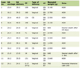 Click to view | Table 1. GC Incidence in Patients From Families With HBOC Based on Genetic Testinga |
A novel treatment method was examined using cancer genome panel testing in 47,211 Japanese patients with advanced-stage/metastatic GC. Overall, 2,417 of 2,489 patients (97.11%) with advanced stage/metastatic GC were infected with H. pylori based on cancer genome panel testing. Of the 173 and 41 patients with GC from families with and without HBOC, 161 (93.06%) and 39 (95.12%) had H. pylori infection, respectively. The incidences of H. pylori infection in patients with GC who were members of families with versus without HBOC were comparable.
These data suggested that individuals who carry gBRCA1/2 PV but are not infected with H. pylori do not have a higher incidence of GC. Compared to individuals with gBRCA1/2 wt, those with gBRCA1/2 PVs have a higher incidence of GC owing to H. pylori infection [5, 7, 8]. Therefore, H. pylori eradication using antibiotics can reduce the incidence of GC even in gBRCA1/2 PV carriers.
In families with HBOC, the risk of developing stomach cancer in men is higher than that in women. However, among individuals from families without HBOC, the risk of developing stomach cancer is roughly equal between the sexes. Therefore, we examined the cumulative risk of stomach cancer affliction by the age of 80 years in each cohort and found it to be 2.71% and 0.91% among men and women in the HBOC group, respectively, whereas these rates were 0.46% and 0.40% in the non-HBOC group, respectively (Fig. 1). The lifetime risk of developing stomach cancer among men is reported to be two- to threefold higher than that in women; this is thought to be owing to the fact that the cumulative rate of stomach cancer among men from HBOC families is approximately three times higher than that among women.
 Click for large image | Figure 1. Cumulative risk of gastric cancer in individuals up to 80 years of age according to family hereditary breast and ovarian cancer (HBOC) statuses. The cumulative risks of gastric cancer were estimated for men and women who underwent National Cancer Genome profiling. *P < 0.001, **Not significant. |
Differences in cancer risk according to sex in Gan mice
In families with HBOC, the cumulative risk of being diagnosed with GC by 80 years of age was 2.71% in men and 0.91% in women (Fig. 1). We used genetically modified mice (Gan mice) that spontaneously develop GC to investigate the reason for this discrepancy between the sexes. Gastric mucosal tissue from C57BL/6 wt mice is shown in panels E1 and E4 of Figure 2a while that from Gan mice is shown in panels E2, E3, E5, and E6 of Figure 2a. Compared to male Gan mice of the same age (Fig. 2a: panels E5, E6), the gastric mucosal tissue of female Gan mice clearly had fewer atypical and highly atypical cells (Fig. 2a: panels E2, E3). Only a few inflammatory cells were observed in the gastric mucosal tissue of 8-month-old female Gan mice while numerous such cells were observed in the mucosal tissue of 12-month-old female Gan mice (Fig. 2b: panels E12, E13). On the other hand, in male Gan mice, high-grade nuclear and cellular atypia was observed in the gastric cancer mucosal tissue (Fig. 2b: panels E12, E13). Hence, endogenous factors expressed in males versus females may act on gastric mucosal cells to suppress carcinogenesis.
 Click for large image | Figure 2. Comparison of gastric tissue samples. Compared to male Gan mice, female Gan mice showed fewer inflammatory and highly dysplastic (adenocarcinoma) cells in their gastric mucus tissues. (a) Panels 1 and 4 show the pathological findings of normal gastric mucosal tissue from a C57BLACK/6 wild-type mouse. Genetically engineered Gan mice exhibit to spontaneously develop gastritis and gastric cancer (panels E2, E3, E5, E6). Compared to the gastric mucosal tissue of male Gan mice of the same age (E5, E6), the gastric mucosal tissue of female Gan mice clearly had fewer atypical and highly atypical cells (E2, E3). (b) Only a few inflammatory cells were observed in the gastric mucosal tissues of 8-month-old Gan mice (panels: E11, E12). However, many inflammatory cells were observed in the mucosal tissues of 12-month-old female Gan mice (panels E12, E13). Scale bar: 100 mm. |
Development of atypical cells in the gastric mucosal tissue of ovariectomized female C57BL/6 mice
Compared to the mucosal tissues of female Gan mice ≤ 12 months of age, numerous highly dysplastic and cancer cells were observed in the mucosal tissues of male counterparts (Fig. 2). Sex-based differences have been observed in the incidence rates of hepatocellular carcinoma and pancreatic cancer, and previous studies suggested that this is owing to sex hormones (i.e., the anti-tumor effects of female hormones such as estrogen and progesterone versus the tumor-enhancing effects of testosterone). However, no clear changes in tumor formation rates have been demonstrated when sex hormones are administered to individuals.
The BRCA2 protein, a transcription product of BRCA2, is a tumor suppressor that forms a large complex in the nucleus with numerous other tumor suppressor factors and is involved in homology-mediated gene repair. We investigated whether pathogenic mutations in BRCA2 result in genetic instability that can ultimately lead to breast and ovarian cancer induction by estrogen and progesterone (Fig. 3a). In two cultured gastric adenocarcinoma cells (AGS cells and MKN28 cells), the expression of the BRCA2 gene and protein was induced in an estrogen concentration-dependent manner (Fig. 3a). In cultured breast cancer (MCF7) and cervical cancer (HeLa) cells, BRCA2 gene and protein expression also was induced in an estrogen concentration-dependent manner (Supplementary Material 2, wjon.elmerpub.com). However, no BRCA2 protein expression was observed in cells stimulated with progesterone (Supplementary Material 3, wjon.elmerpub.com). We previously showed that the incidence of GC is reduced in Gan BRCA2 transgenic mice (which constitutively express BRCA2) compared to Gan mice (Supplementary Material 4, wjon.elmerpub.com). In other words, it is feasible that estrogen-induced BRCA2 acts as an anti-tumor agent in the mucosal epithelium of female Gan mice, resulting in a lower incidence of malignant tumors compared to that in male Gan mice (Supplementary Material 4, wjon.elmerpub.com). Next, we investigated the incidence of GC in C57BL/6, Gan, and Gan BRCA2 mice. No dysplastic cells were detected in the gastric mucosa of normal male and female C57BL/6 mice; however, low-grade dysplastic cells were observed in the gastric mucosa of female C57BL/6-ovariectomy mice in which the estrogen-secreting ovaries had been removed; neither high-grade dysplastic cells nor cancer cells were detected (Fig. 3c; Supplementary Material 5, wjon.elmerpub.com).
 Click for large image | Figure 3. BRCA2 is induced by estrogen stimulation. (a) We investigated whether pathogenic mutations in the BRCA2 gene result in genetic instability that can ultimately lead to breast and ovarian cancer induction by estrogen. BRCA2 protein expression was induced in an estrogen concentration-dependent manner in cultured gastric adenocarcinoma cells (AGS cells and MKN28 cells). (b) Schematic representing BRCA2 gene expression induced by estrogen stimulation. Notably, no induction of BRCA2 protein was observed in progesterone-stimulated MCF7 and HeLa cells. (c) Pathological analysis was performed on the gastric tissues of mice carrying certain genetic modifications that were sacrificed at 14 months of age. Based on evidence obtained from our reserch that estrogen-induced BRCA2 may protect against tumor development in the mucosal epithelium of female Gan mice, we investigated the incidence of gastric cancer in C57BL/6, Gan, and Gan BRCA2 mice. No dysplastic cells were detected in the gastric mucosa of normal male and female C57BL/6 mice. Therefore, for female C57BL/6 mice (ovariectomized mice), the number of mice in which only cells exhibiting mild. The asterisk: However, in the gastric mucosa of female C57BL/6-ovariectomy mice in which the estrogen-secreting ovaries had been removed, only low-grade dysplastic cells were observed; neither high-grade dysplastic cells nor cancer cells were detected, therefore, for female C57BL/6 mice (ovariectomized mice), the number of mice in which only cells exhibiting mild dysplasia were observed in the gastric mucosa is reported. |
High-grade dysplastic and cancer cells were observed in the gastric mucosal tissues of both female and male Gan mice (Fig. 3c). Furthermore, the tumor incidence rate was higher in the gastric mucosal tissue of female Gan-ovariectomy mice (Fig. 3c; Supplementary Material 5, wjon.elmerpub.com). These findings suggest that the absence of estrogen secretion following ovariectomy attenuates BRCA2 expression in the gastric mucosal tissue of Gan-ovariectomy mice, resulting in the emergence of numerous high-grade dysplastic and cancer cells.
Value of BRCA2 as a prognostic marker in GC
Studies using genetically modified mice demonstrated that BRCA2 expression reduces the risk of developing GC. We therefore investigated the relationship between BRCA2 expression and the prognosis of patients with GC using data collected from a cancer genome medicine clinical trial conducted at Kyoto University Hospital. Compared to normal gastric mucosal tissue in men, strong expression of estrogen receptor α and BRCA2 was observed in normal gastric mucosal tissue in women. Furthermore, estrogen blood concentrations in women (during ovulation and luteal phase) were significantly higher than those in men (Supplementary Material 6, wjon.elmerpub.com). Compared to men, the concentration of estrogen in the blood of women (during ovulation and the luteal phase) was markedly higher (Supplementary Material 6, wjon.elmerpub.com). We examined the expression status of BRCA2 in normal gastric mucosal tissue (407 cases) and GC mucosal tissue (346 cases) using immunohistochemical staining. We found that BRCA2 expression was strong in most of the normal gastric mucosal tissue samples (Fig. 4a, left panel) but was weak or negative in most of the GC mucosal tissue samples (Fig. 4a, right panel).
 Click for large image | Figure 4. The value of BRCA2 expression status as a prognosis marker. (a) We examined the expression status of BRCA2 in normal gastric mucosal tissue (407 cases) and gastric cancer mucosal tissue (346 cases) using immunohistochemical staining. BRCA2 expression was high in normal gastric mucosal tissue but weak or negative in the mucosal tissue of many gastric cancers. Scale bar: 30 mm. (b) The Mantle 2 color imaging system was used to quantify the expression intensity of BRCA2 in various gastric mucosal tissues. A scatter plot of BRCA2 expression levels revealed a cutoff value of 3.0 to separate high- versus low-expression groups. (c) Patients with gastric cancer were divided into two groups based on BRCA2 expression levels in their resected gastric mucosal tissue: low-BRCA22 (235 cases) and high-BRCA2 expression (111 cases). Kaplan–Meier survival curves revealed longer survival rates in the high-BRCA2 group than in the low BRCA2 group. (d) The median overall survival rates (50%) were 2.1 and 3.7 years in the BRCA2 low and BRCA2 high groups. The 5-year survival rates in the BRCA2 low and BRCA2 high groups were 34.2% and 41.2%. Please refer to Supplementary Material 7 (wjon.elmerpub.com) for details regarding the gastric cancer stage and other information for each patient who participated in this clinical study. F/M/total cases: female cases/male cases/total cases. |
Next, we divided these patients into two groups: those with low BRCA2 expression in GC mucosal tissue (235 cases) and those with high BRCA2 expression (111 cases). Using the Kaplan–Meier method, we found that the median overall survival rates (50%) were 2.1 and 3.7 years in the BRCA2 low and BRCA2 high groups. The 5-year survival rates in the BRCA2 low and BRCA2 high groups were 34.2% and 41.2% (Fig. 4c, d). In this clinical study, women with GC who expressed high levels of BRCA2 had a better prognosis compared to other cohorts, suggesting that BRCA2 may be a prognostic marker for GC (Fig. 4c, d; Supplementary Material 7, wjon.elmerpub.com).
| Discussion | ▴Top |
The incidence of GC increases after 50 years of age, particularly among men; however, the reason for the higher incidence rate in men than in women remains unclear. Older studies did not consider the individuals’ genetic backgrounds. We previously found that the risk of GC is indeed influenced by an individual’s genetic background, including whether gBRCA1/2 PVs are present, and is significantly increased by H. pylori infection. In 1994, the World Health Organization designated H. pylori as a group I (definite) carcinogen similar to tobacco [9]. Momozawa et al and Hayashi et al have shown that pathogenic mutations in BRCA1 or BRCA2 have a significant impact on GC development to an extent similar to infection with CagA positive-H. pylori. Individual genetic backgrounds, especially the presence of homologous recombination deficiency genes including BRCA1/2 PVs, ATM, BARD1, BRCA1, BRCA2, BRIP1, CHEK2, PALB2, RAD51C, and RAD51D have a significant impact on GC development caused by H. pylori infection [4, 7, 10]. Kanno et al provided a clinical explanation for the impact of BRCA1/2 PVs on the development of GC [11]. We used genetically modified mice as well as resected human tissues to investigate the sex-based differences in the risk of developing GC using molecular pathological techniques. We found that estrogen stimulation induces BRCA2 expression, which in turn reduces the risk of GC in female mice compared to male mice. Clinical studies have revealed that patients with weaker BRCA2 expression in GC tissues tend to have shorter survival rates. The higher incidence of GC in men may therefore be due to the underlying cause of hereditary GC.
Several viruses are known to cause cancers. Human papillomavirus (HPV) is considered the main cause of cervical cancer [10, 12] and is spread through sexual contact; therefore, anyone with sexual contact is at risk of HPV infection. Whereas a majority of individuals within a population may be infected with HPV, the virus is often eliminated by the person’s immune system. In rare instances, HPV can become chronic and can cause cervical, penile, and laryngeal cancer. Hepatitis B (HBV) and C viruses (HCV) infect the liver cells, causing chronic hepatitis and cirrhosis [13, 14]. Furthermore, hepatocellular carcinoma (HCC) often develops from chronic cirrhosis caused by HBV or HCV infection. HBV is primarily transmitted through blood, whereas HCV is chiefly transmitted through needles and other blood-contacting devices. However, no study to date has investigated the relationship between these viral infections and homologous recombination deficiency, including BRCA1/2 PVs, during carcinogenesis. CagA secreted by H. pylori binds to BRCA1/2 wt, BRCA1/2 PVs and induced the infected cells from undergoing apoptosis [15, 16]. Virus-derived proteins bind to BRCA1/2 PVs and induce tumorigenesis. Previous studies have generally not revealed any association between GC and most physiological and reproductive factors. However, our current findings revealed that estrogen stimulation strongly induces BRCA2 expression in female mice, which in turn suppresses GC. Future research will shed light on the roles of virus-derived factors and their complexes with BRCA1/2 PVs in carcinogenesis. While vaccination may be an effective method against viral infections, the development of an effective vaccines remains challenging.
There are some other cancers whose incidence rates differ according to sex. The incidence of rate is approximately twice as high in men as in women; it begins to increase in men in their 40s and women in their 50s and is most common among individuals aged > 80 years. [17]. HCC is caused by chronic hepatitis and cirrhosis due to HBV and HCV infection, although the incidence rate has been decreasing owing to thorough infection prevention measures and advances in antiviral therapy [18]. Conversely, there has been an increase in the incidence of HCC caused by alcohol-related liver disease and metabolic dysfunction associated with steatotic liver disease, which are not virus-related [19] but stem from lifestyle factors such as alcohol consumption, obesity, diabetes, hypertension, and dyslipidemia. However, the molecular and biological mechanisms that explain the sex differences in the incidence of HCC remain unclear.
Clinical studies conducted to date have shown that the incidence of GC is higher in individuals or families exhibiting functional loss mutations or a lack of expression in the BRCA1 or BRCA2 genes, compared to the incidence in the general population [4, 5, 7]. Accordingly, families in which pathogenic germline BRCA1/2 variants are identified are classified as having HBOC syndrome and exhibit a high incidence of GC [7]. Furthermore, within HBOC families, the incidence of GC is lower in women than in men.
Japanese medical staff are conducting research to clarify the relationship between various lifestyle habits and diseases such as cancer, stroke, and myocardial infarction, with the aim of contributing to the prevention of lifestyle-related diseases among the Japanese population. Since the incidence of stomach cancer is about 1/2 to 1/3 lower in women than in men, it is speculated that some female-specific factors, such as menstruation or reproduction (female-related factors), may be associated with stomach cancer [20]. In clinical studies conducted in Japan to date, participants have been grouped based on reproductive factors such as age at menarche and number of births, and the association between these female-related factors and the risk of developing GC has been investigated. It has been shown that individuals who experienced menarche at an early age (12 years or younger) have a 0.65 times lower risk of developing GC compared to those who experienced it at a later age (15 years or older) [20]. This trend remained consistent regardless of the histological type of the GC (differentiated or undifferentiated) or the location of the tumor within the stomach [20]. The regulation of BRCA2 expression is associated with the binding of USF, Elf1, and NF-κB transcription factors to the BRCA2 promoter. The mechanism of E2-induced BRCA2 promoter activation in breast cancer cells (MCF7) was analyzed [21]. As a result, two Sp1 sites responsive to E2-ER complex activity were identified [21].
While hormone replacement therapy (HRT) is widely used to alleviate menopausal symptoms, the association between HRT use and GC remains unclear. Subgroup analyses based on HRT formulation type revealed a reduced risk of GC for both estrogen-only therapy (pooled relative risk (RR): 0.63; 95% confidence interval (CI): 0.51–0.77; I2 = 0%) and combined estrogen-progestin therapy (pooled RR: 0.70; 95% CI: 0.57–0.87; I2 = 0%) compared to non-use [22]. In systematic reviews and meta-analyses, HRT use was associated with a reduced risk of GC, regardless of the formulation type [22].
Through a large-scale evaluation of patients with different clinical and demographic characteristics, we identified genes associated with an increased risk of GC in families with HBOC and BRCA1/2 PVs. H. pylori infection increased the risk of GC associated with germline BRCA1/2 PVs in patients from families with HBOC. Our results suggest that the evaluation and eradication of H. pylori infection may be beneficial for individuals with HBOC and gBRCA1/2 PVs.
| Supplementary Material | ▴Top |
Suppl 1. Materials and methods.
Suppl 2. Progesterone stimulation affects BRCA2 expression.
Suppl 3. Progesterone stimulation did not affect BRCA2 expression.
Suppl 4. (A) The mRNA expression of BRAC1 or BRCA2 in GantgBRCA1 or GantgBRCA2 transgenic mice. (B) The of BRAC1 or BRCA2 transgene expression in GantgBRCA1 or GantgBRCA2 transgenic animals.
Suppl 5. No dysplastic cells were detected in the gastric mucosa of normal male and female C57BL/6 mice; however, low-grade dysplastic cells were observed in the gastric mucosa of female C57BL/6-ovariectomy mice in which the estrogen-secreting ovaries had been removed; neither high-grade dysplastic cells nor cancer cells were detected.
Suppl 6. Possibility of estrogen-induced BRCA2 expression in gastric mucosal tissue cells.
Suppl 7. Number of samples (total 346 cases).
Acknowledgments
The authors thank all medical staff for clinical research at the National Hospital Organization Kyoto Medical Center.
Financial Disclosure
The costs of conducting our clinical research are covered by the following research grants: Japan Society for Promoting Science for TH (No. 19K09840), START-program Japan Science and Technology Agency (JST) for TH (No. STSC20001), National Hospital Organization Multicenter Clinical Study for TH (Grant No. 2019-Cancer in general-02), and Japan Agency for Medical Research and Development (AMED) (No. 22ym0126802j0001), Tokyo, Japan. These research funds were provided by Japanese government research institutes, and therefore the funders were not involved in the establishment of the clinical research program, the collection and analysis of data, the public dissemination of research findings, or the preparation of the manuscript.
Conflict of Interest
The authors declare no potential conflict of interest. The founders had no role in study design, data collection and analysis, decision to publish, or preparation of the manuscript.
Informed Consent
We received informed consent forms from patients participating in the clinical research.
Author Contributions
TH is responsible for research design and writing manuscript. TH and IK were involved in the literature search, study design, data collection, and data interpretation. IK reviewed the manuscript. TH and IK are responsible for final content. All authors have read and approved the final version of the manuscript.
Data Availability
The details of the materials and methods are indicated in the online supplementary file. The data are stored in a repository external to the journal's homepage and have been made publicly available; the data are also publicly accessible on various websites, and details regarding data availability are provided in the first paragraph of the Results section. Information from this clinical study and the associated transparency statement in the medical journal article are available online.
| References | ▴Top |
- Sugano K, Suzuki C, Ota M, Iwakiri R. Gastric cancer risk after Helicobacter pylori eradication in gastritis and peptic ulcer: a retrospective cohort study in Japan. BMC Gastroenterol. 2025;25(1):463.
doi pubmed - Hatakeyama M. Helicobacter pylori CagA and gastric cancer: a paradigm for hit-and-run carcinogenesis. Cell Host Microbe. 2014;15(3):306-316.
doi pubmed - Floch P, Laur AM, Korolik V, Chrisment D, Cappellen D, Idrissi Y, Dubus P, et al. Characterisation of inflammatory processes in Helicobacter pylori-induced gastric lymphomagenesis in a mouse model. Oncotarget. 2015;6(33):34525-34536.
doi pubmed - Momozawa Y, Sasai R, Usui Y, Shiraishi K, Iwasaki Y, Taniyama Y, Parsons MT, et al. Expansion of cancer risk profile for BRCA1 and BRCA2 pathogenic variants. JAMA Oncol. 2022;8(6):871-878.
doi pubmed - Usui Y, Taniyama Y, Endo M, Koyanagi YN, Kasugai Y, Oze I, Ito H, et al. Helicobacter pylori, homologous-recombination genes, and gastric cancer. N Engl J Med. 2023;388(13):1181-1190.
doi pubmed - http://n2t.net/addgene:16246.
- Hayashi T, Sano K, Okada M, Ura T, Konishi I. Hereditary gastric cancer is linked with hereditary breast and ovarian cancer. World J Oncol. 2024;15(4):722-730.
doi pubmed - Hayashi T, Sano K, Okada M, Muto M, Konishi I. Efficacy and tolerability of olaparib plus paclitaxel in patients with gastric cancer associated with hereditary breast and ovarian cancer. Curr Oncol. 2024;31(11):6723-6734.
doi pubmed - De Luca A, Iaquinto G. Helicobacter pylori and gastric diseases: a dangerous association. Cancer Lett. 2004;213(1):1-10.
doi pubmed - Pilie PG, Tang C, Mills GB, Yap TA. State-of-the-art strategies for targeting the DNA damage response in cancer. Nat Rev Clin Oncol. 2019;16(2):81-104.
doi pubmed - Kanno M, Fusegi A, Miyazaki N, Ozawa R, Netsu S, Aoki Y, Omi M, et al. Surgical timing and survival in advanced high-grade serous ovarian cancer in the PARP inhibitor era. Cancers (Basel). 2026;18(2):245.
doi pubmed - Naucler P, Ryd W, Tornberg S, Strand A, Wadell G, Elfgren K, Radberg T, et al. Human papillomavirus and Papanicolaou tests to screen for cervical cancer. N Engl J Med. 2007;357(16):1589-1597.
doi pubmed - D'Errico A, Grigioni WF, Fiorentino M, Baccarini P, Lamas E, De Mitri S, Gozzetti G, et al. Expression of insulin-like growth factor II (IGF-II) in human hepatocellular carcinomas: an immunohistochemical study. Pathol Int. 1994;44(2):131-137.
doi pubmed - Ambrosch A, Konig W. [Characteristics of the hepatitis C virus and viral predictors of therapeutic response]. Med Klin (Munich). 1999;94(11):626-632.
doi pubmed - Hatakeyama M. Oncogenic mechanisms of the Helicobacter pylori CagA protein. Nat Rev Cancer. 2004;4(9):688-694.
doi pubmed - Imai S, Ooki T, Murata-Kamiya N, Komura D, Tahmina K, Wu W, Takahashi-Kanemitsu A, et al. Helicobacter pylori CagA elicits BRCAness to induce genome instability that may underlie bacterial gastric carcinogenesis. Cell Host Microbe. 2021;29(6):941-958.e910.
doi pubmed - Bracco C, Gallarate M, Badinella Martini M, Magnino C, D'Agnano S, Canta R, Racca G, et al. Epidemiology, therapy and outcome of hepatocellular carcinoma between 2010 and 2019 in Piedmont, Italy. World J Gastrointest Oncol. 2024;16(3):761-772.
doi pubmed - Colombo M, Lleo A. The impact of antiviral therapy on hepatocellular carcinoma epidemiology. Hepat Oncol. 2018;5(1):HEP03.
doi pubmed - Sandella R, Rinella ME. Metabolic and Alcohol-Associated: Diagnosis and Management. Clin Liver Dis. 2026;30(1):167-183.
doi pubmed - Persson C, Inoue M, Sasazuki S, Kurahashi N, Iwasaki M, Ye W, Tsugane S, et al. Female reproductive factors and the risk of gastric cancer in a large-scale population-based cohort study in Japan (JPHC study). Eur J Cancer Prev. 2008;17(4):345-353.
doi pubmed - Li PC, Zhu YF, Cao WM, Li B. ER-positive and BRCA2-mutated breast cancer: a literature review. Eur J Med Res. 2024;29(1):30.
doi pubmed - Jang YC, Leung CY, Huang HL. Association of hormone replacement therapy with risk of gastric cancer: a systematic review and meta-analysis. Sci Rep. 2022;12(1):12997.
doi pubmed
This article is distributed under the terms of the Creative Commons Attribution 4.0 International License (CC BY 4.0), which permits unrestricted use, distribution, and reproduction in any medium, including commercial use, provided the original work is properly cited.
World Journal of Oncology is published by Elmer Press Inc.