| World Journal of Oncology, ISSN 1920-4531 print, 1920-454X online, Open Access |
| Article copyright, the authors; Journal compilation copyright, World J Oncol and Elmer Press Inc |
| Journal website https://wjon.elmerpub.com |
Original Article
Volume 17, Number 6, December 2026, pages 830-843

The Development of Breast Apocrine Carcinoma Score That Distinguishes Breast and Cutaneous Apocrine Carcinomas
Kei Kawashimaa, b, c, Akimitsu Yamadab, c, Eleanor A. Fallona, d, Ryo Fujitab, c, Mahato Sasamotob, c, Masanori Oshib, c, Kazutaka Naruie, Hideyuki Ishikawaf, Jotaro Haradag, Yukie Yamaguchif, Satoshi Fujiig, h, Takashi Ishikawac, i, Itaru Endob, Kazuaki Takabea, b, d, i, j, k, l
aDepartment of Surgical Oncology, Roswell Park Comprehensive Cancer Center, Buffalo, NY 14263, USA
bDepartment of Gastroenterological Surgery, Yokohama City University Graduate School of Medicine, Kanagawa, Japan
cDepartment of Breast and Endocrine Surgery, Yokohama City University Graduate School of Medicine, Kanagawa, Japan
dDepartment of Surgery, University at Buffalo Jacobs School of Medicine and Biomedical Sciences, The State University of New York, Buffalo, NY, USA
eDepartment of Breast and Thyroid Surgery, Yokohama City University Medical Center, Yokohama, Kanagawa, Japan
fDepartment of Environmental Immuno-Dermatology, Yokohama City University Graduate School of Medicine, Yokohama, Japan
gDepartment of Molecular Pathology, Yokohama City University Graduate School of Medicine, Yokohama, Japan
hDepartment of Pathology, Yokohama City University Hospital, Yokohama, Japan
iDepartment of Breast Surgery and Oncology, Tokyo Medical University, Tokyo, Japan
jDivision of Digestive and General Surgery, Niigata University Graduate School of Medical and Dental Sciences, Niigata, Japan
kDepartment of Breast Surgery, Fukushima Medical University, Fukushima, Japan
lCorresponding Author: Kazuaki Takabe, Department of Surgical Oncology, Roswell Park Comprehensive Cancer Center, Buffalo, NY 14263, USA
Manuscript submitted August 27, 2026, accepted September 23, 2026, published online November 11, 2026
Short title: Breast vs Cutaneous Apocrine Carcinoma
doi: https://doi.org/10.14740/wjon2869
| Abstract | ▴Top |
Background: Apocrine carcinoma (AC) is a rare cancer that arises either from cutaneous apocrine sweat glands (cutaneous AC) or as breast carcinoma with apocrine differentiation (breast AC). Differentiation between them is clinically critical because their management and outcomes differ significantly: breast AC is treated according to established breast cancer therapies, whereas cutaneous AC lacks standardized systemic treatment and often carries a poorer prognosis. A diagnostic challenge arises when AC occurs in the axilla, where both entities may arise, given their highly similar histology.
Methods: In this study, we performed a retrospective multi-cohort transcriptomic analysis integrating institutional, multi-institutional, and publicly available datasets to characterize the molecular differences between breast and cutaneous ACs and developed a transcriptomic classifier using differentially expressed genes and a least absolute shrinkage and selection operator (LASSO) logistic regression model for tissue-of-origin classification.
Results: Transcriptomic profiling revealed that breast ACs exhibited higher steroid hormone–related signaling and lower proliferative activity compared with cutaneous ACs, while immune-related features were largely comparable between the two tumor types. These findings suggest that, despite shared apocrine morphology, breast and cutaneous ACs retain distinct biological characteristics reflective of their tissue of origin. Building on this, we further developed a transcriptomic classifier, the Breast Apocrine Carcinoma (BAC) score, which distinguished breast and cutaneous ACs, suggesting that these tumors retain distinct tissue-of-origin transcriptional signatures despite shared apocrine differentiation. Furthermore, application of the BAC score to tumors with unknown primary origin in the axillary region enabled estimation of tissue origin, highlighting its potential clinical utility in diagnostically challenging cases.
Conclusions: Breast and cutaneous ACs exhibit distinct biological features despite shared apocrine morphology. The BAC score showed promising discriminatory ability for tumors of breast and cutaneous origin, including within AC cohorts, highlighting the potential utility of transcriptomic profiling for improving their classification and diagnostic evaluation.
Keywords: Breast apocrine carcinoma; Cutaneous apocrine carcinoma; Differentiation; Transcriptome; Discrimination score
| Introduction | ▴Top |
Apocrine glands are specialized sweat glands in the skin that secrete lipid-rich sweat and are predominantly distributed in the axilla [1]. Tumors derived from these glands exhibit apocrine differentiation and are collectively referred to as cutaneous apocrine carcinomas (AC) [2], which is a rare adnexal malignancy of the skin. Incidence of cutaneous AC ranges from approximately 0.0049 to 0.23 per 100,000 persons per year across different populations and databases, and no significant temporal change was observed in recent epidemiologic analyses [2, 3]. Interestingly, AC can also occur in the breast. The mammary glands in breast and apocrine glands share common embryological origins [4–6], and apocrine differentiation is commonly observed in breast tissue [7]. As a matter of fact, autopsy studies have shown that 50–85% of normal breast tissue exhibits some degree of apocrine morphology [8–10]. When apocrine morphology occupies the majority of cancer cells, the tumor is classified as breast AC [11], which accounts for approximately 0.4–4% of all breast cancers [12, 13].
Differentiation between breast and cutaneous AC is clinically critical, as treatment strategies and prognoses differ substantially. Breast AC is generally managed according to therapeutic approaches for invasive breast carcinoma of no special type (NST), with therapeutic decisions being guided by tumor subtype [12]. Breast AC has been reported to show relatively favorable clinical outcomes compared with non-apocrine breast cancers, particularly within triple-negative breast cancer (TNBC) [13–15], which otherwise is considered the most aggressive subtype with the worst outcomes. Building upon this, we recently reported that the presence of apocrine morphology in breast cancer is associated with distinct estrogen receptor (ER)-dependent biological and clinical features [16]. In contrast, cutaneous AC is primarily treated with surgical excision, and there is currently no established systemic therapy [17]. The prognosis of cutaneous AC has been reported to be poor, with median survival ranging from approximately 33 to 55 months depending on nodal status [18].
Although critically important, distinguishing breast and cutaneous ACs has long been a clinical challenge [18] particularly in the axilla because both subtypes can occur in this anatomic region [2, 19–21]. Ectopic breast tissue most commonly occurs in the axilla [22] and is present in approximately 1–6% of the population [23, 24]. Although rare, this tissue can give rise to ectopic breast cancer, which accounts for approximately 0.3–0.6% of all breast cancers [20, 21]. Conversely, cutaneous ACs frequently occur in the axilla as it is rich in apocrine sweat glands [2, 19]. Histologically, breast and cutaneous ACs share highly similar morphological features, including abundant eosinophilic cytoplasm and apocrine differentiation, making distinction based on morphology alone difficult [18]. Immunohistochemical markers commonly used in clinical practice, including mammaglobin and gross cystic disease fluid protein (GCDFP), also show substantial overlap between the two entities, reflecting shared apocrine differentiation [6, 18]. Conversely, markers such as p63 and CK5/6, which may favor a primary cutaneous adnexal origin, are not entirely specific. Therefore, because morphology alone is insufficient and immunohistochemical profiles substantially overlap, distinguishing primary cutaneous AC from cutaneous metastasis of breast AC can remain difficult even when clinical and pathological findings are considered together. To this end, there is an unmet need for reliable approaches to distinguish breast AC from cutaneous AC to ensure appropriate treatment selection.
To date, comprehensive molecular comparisons between breast and cutaneous ACs have not been systematically performed. In particular, transcriptomic differences between the two entities remain largely understudied. Given this unmet clinical need, we conducted a retrospective multi-cohort study to comprehensively characterize the clinical and molecular distinctions between cutaneous and breast ACs. Furthermore, we developed a novel transcriptomic scoring system, the Breast Apocrine Carcinoma (BAC) score, to facilitate their differentiation, which may serve as a potential tool to distinguish cutaneous and breast ACs.
| Material and Methods | ▴Top |
Study design and patient cohort
This retrospective multi-cohort study included a multi-institutional cohort from the Oncology Research Information Exchange Network (ORIEN) and an institutional cohort from Yokohama City University (YCU) hospital, and publicly available transcriptomic datasets. The ORIEN cohort included patients diagnosed with breast cancer (n = 1,797) or skin cancer (n = 331). Among these cases, four cases of breast AC and two cases of cutaneous AC were identified and defined as the ORIEN apocrine subset. The remaining breast and skin cancer cases were defined as the ORIEN non-apocrine subset and were used as the training cohort for transcriptomic score development. Deidentified clinical, pathological, and sequencing data, including RNA sequencing and whole-exome sequencing (WES) data, were obtained from the ORIEN Avatar program under Institutional Review Board (IRB)-approved protocols at Roswell Park Comprehensive Cancer Center (approved number: BDR 198425). The YCU cohort consisted of patients diagnosed with breast AC (n = 4) or primary cutaneous AC (n = 2) at the Departments of Breast Surgery and Dermatology, YCU hospital, between 2017 and 2023. These cases were defined as the YCU reference subset. In addition, ACs arising in the axillary region with unknown primary origin (breast versus cutaneous) were included and defined as the YCU unknown-primary subset (n = 4). The study was approved by the IRB of YCU (approved number: F240200004). Clinical and pathological data were collected retrospectively from electronic medical records. RNA was extracted from formalin-fixed paraffin-embedded (FFPE) core needle biopsy specimens obtained at diagnosis or from residual FFPE tissue from surgical resections.
Publicly available transcriptomic datasets were accessed for additional analyses. Transcriptomic and clinical data for breast cancer and skin cancer from The Cancer Genome Atlas (TCGA) [25], the Cancer Cell Line Encyclopedia (CCLE) [26] and MET500 [27] were downloaded from cBioPortal and UCSC Xena and analyzed. All datasets were accessed and downloaded in October 2025.
Sequencing methods
For the YCU cohort, FFPE specimens were sectioned at a thickness of 10 µm and stained with hematoxylin and eosin (H&E). Tumor regions were selectively isolated using laser microdissection (LMD7000, Leica Microsystems). Total RNA was extracted using the RNeasy FFPE Kit (Qiagen, Venlo, Netherlands) according to the manufacturer’s instructions. RNA sequencing was performed by RIKEN Genesis Co., Ltd. For the ORIEN cohort, transcriptome and WES data were generated through the ORIEN Avatar program using the standardized Total Cancer Care (TCC) protocol as previously described [28].
Transcriptomic analysis
Principal component analysis (PCA) was conducted using the gene expression values to visualize global transcriptomic differences between breast and cutaneous ACs. Differentially expressed gene (DEG) analysis was performed using the limma package in R. To evaluate pathway activity at the individual sample level, single-sample gene set enrichment analysis (ssGSEA) scores were calculated using curated gene sets from the Molecular Signatures Database (MSigDB) [29] using fgsea package. These scores represent the relative enrichment of predefined biological pathways in each sample. The relative abundance of immune cell populations in the tumor microenvironment was estimated using the xCell algorithm [30]. Cytolytic activity (CYT) scores were calculated as the geometric mean of granzyme A (GZMA) and perforin (PRF1) expression, as described by Rooney et al [31]. Gene expression levels of selected individual genes were analyzed to evaluate the expression patterns of hormone receptor–related and other biologically relevant markers. Prediction Analysis of Microarray 50 (PAM50) intrinsic subtypes were inferred using the genefu package [32] in R.
Development of a transcriptomic score to distinguish breast and cutaneous tumors
Because ACs are extremely rare and the available sample size was limited, a transcriptomic classification model to distinguish breast and cutaneous origins was developed using a larger cohort of non-apocrine breast and skin cancers from the ORIEN cohort non-apocrine subset as the training cohort. The model generated a transcriptomic score, termed the BAC score. Genes with low variability were filtered based on the median absolute deviation (MAD), retaining genes above the 25th percentile of MAD calculated within the training samples. Gene expression values were standardized using Z-score normalization based on the mean and standard deviation calculated from the training cohort. To incorporate biologically informative genes, differential expression analysis between breast and skin tumors in the training cohort was performed using the limma package, and the top DEGs were assigned reduced penalty weights (penalty factor = 0.5) in the model to encourage their selection. A least absolute shrinkage and selection operator (LASSO) logistic regression model was then fitted to predict breast versus skin origin. Class weights were applied to account for imbalance in sample sizes between the two tumor types. The regularization parameter (λ) was selected using 10-fold cross-validation based on the one-standard-error rule (λ1se). The resulting model generated a transcriptomic score (BAC score), defined as a linear predictor:
The BAC score was developed by leveraging multiple independent breast and skin cancer cohorts (TCGA, breast n = 1,081, cutaneous n = 470; CCLE, breast n = 77, cutaneous n = 119; MET500, breast n = 159, cutaneous n = 139), as well as the ORIEN apocrine subset (breast AC, n = 4; cutaneous AC, n = 2) and the YCU reference subset (breast AC, n = 4; cutaneous AC, n = 2). Within the YCU reference subset, the optimal cutoff value for the BAC score was determined using the Youden index. Given the small number of cases in this subset, this cutoff was considered exploratory rather than an established diagnostic threshold and requires validation in an independent cohort. This cutoff was subsequently applied to the YCU unknown-primary subset to assign predicted labels. Transcriptomic features of these predicted cases were then compared with those of the YCU reference subset.
Institutional Review Board approval
This study was approved by the Institutional Review Boards of Roswell Park Comprehensive Cancer Center (BDR 198425) and Yokohama City University (F240200004).
Ethical compliance with human study
This study was conducted in accordance with the ethical standards of the participating institutions and the Declaration of Helsinki.
| Results | ▴Top |
Morphological and clinicopathological characteristics of breast and cutaneous ACs
We first examined the morphological and clinicopathological features of breast and cutaneous ACs. On H&E staining, both tumor types showed highly similar morphological features (Fig. 1a), exhibiting features of apocrine differentiation, including abundant eosinophilic cytoplasm and prominent nucleoli.
 Click for large image | Figure 1. Overview of transcriptomic and mutational features of breast and cutaneous ACs in the ORIEN and YCU cohorts. (a) Representative hematoxylin and eosin (H&E) images of breast and cutaneous ACs in the YCU cohort. Left panels show low-power views and right panels show corresponding high-power views of the same cases. Scale bars are shown in the figure. (b) Principal component analysis (PCA) of transcriptomic profiles in the ORIEN and YCU cohorts. Each point represents one tumor sample; red indicates breast AC and blue indicates cutaneous AC. (c) Volcano plots of differential gene expression between breast and cutaneous ACs in the ORIEN and YCU cohorts. The top 10 upregulated and top 10 downregulated differentially expressed genes are labeled. Red points represent significantly upregulated genes, blue points represent genes that were statistically significant but showed relatively small fold changes, and green points represent genes that did not meet the significance threshold. (d) Oncoplot of recurrent somatic alterations in breast and cutaneous ACs in the ORIEN cohort. Each column represents a tumor sample and each row represents a gene. Alteration types are color-coded as indicated. Alteration frequencies for each gene are shown on the right side of the plot. AC: apocrine carcinoma; ORIEN: Oncology Research Information Exchange Network; YCU: Yokohama City University. |
The clinicopathological characteristics of breast and cutaneous ACs in the ORIEN apocrine subset (breast AC, n = 4; cutaneous AC, n = 2) and the YCU reference subset (breast AC, n = 4; cutaneous AC, n = 2) are summarized in Table 1. The median age of patients with breast AC was 61 years in the ORIEN cohort and 70 years in the YCU cohort, whereas the median age of patients with cutaneous AC was 68 and 50 years, respectively. No consistent age pattern was observed between the two tumor types across cohorts. All breast AC cases occurred in female patients, whereas cutaneous AC occurred in one female and one male patient in the ORIEN cohort, and in two male patients in the YCU cohort. The anatomical sites of cutaneous AC were the scalp (n = 1) and head (n = 1) in the ORIEN cohort, and the scalp (n = 1) and neck (n = 1) in the YCU cohort. In the ORIEN cohort, ER was negative in three cases of breast ACs (75%), and human epidermal growth factor receptor 2 (HER2) was negative in three cases of breast tumors (75%). ER and HER2 status were unavailable for cutaneous tumors. In the YCU cohort, ER was negative in all tumors. HER2 was positive in one breast AC (25%) and in two cutaneous AC (50%).
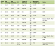 Click to view | Table 1. Clinicopathological Characteristics of Breast and Cutaneous ACs in the ORIEN and YCU Cohorts |
Transcriptomic and genomic overview of ACs
Transcriptomic and genomic analyses were performed for breast and cutaneous AC cases from the ORIEN and YCU cohorts. Tumor purity of RNA-seq samples was estimated using the ESTIMATE package (Supplementary Material 1, wjon.elmerpub.com). The median tumor purity was 0.68 (range, 0.65–0.74) in the ORIEN cohort and 0.90 (range, 0.72–0.94) in the YCU cohort, indicating overall high tumor content in both datasets. PCA based on the top 1,000 highly variable genes demonstrated transcriptomic differences between breast and cutaneous ACs in both cohorts (Fig. 1b). In both cohorts, breast AC cases clustered closely, whereas the two cutaneous AC cases were more widely dispersed in the PCA plot, suggesting greater transcriptomic variability among cutaneous tumors.
To further investigate these transcriptomic differences, DEG analysis was performed between breast and cutaneous ACs in both cohorts (Fig. 1c). Volcano plots with the top 10 DEGs labeled are shown for each cohort. Genes associated with epidermal differentiation, including KRT6A (keratin 6A), SPRR1A (small proline-rich protein 1A), SERPINB3 (serpin family B member 3), and S100A7 (S100 calcium-binding protein A7), were enriched in cutaneous tumors, whereas mammary epithelial lineage markers such as FOXA1 (forkhead box A1), PRLR (prolactin receptor), and ANKRD30A (ankyrin repeat domain 30A) showed higher expression in breast ACs. In addition, several genes previously reported to be associated with apocrine differentiation were identified among the DEGs in the breast tumors of the YCU cohort, including PIP (prolactin-induced protein), ACSM1 (acyl-CoA synthetase medium-chain family member 1), and HMGCS2 (3-hydroxy-3-methylglutaryl-CoA synthase 2).
Somatic mutation analysis was performed in the ORIEN cohort (Fig. 1d). The overall mutational landscape was broadly similar between breast and cutaneous ACs, with no prominent differences observed between the two groups. The most frequently altered genes included MUC16 (mucin 16), TTN (titin), and TP53 (tumor protein p53), along with mutations in genes involved in extracellular matrix organization and cell adhesion, such as FAT4 (FAT atypical cadherin 4) and HSPG2 (heparan sulfate proteoglycan 2).
Breast AC showed enrichment of steroid hormone response-related pathways compared with cutaneous AC
Next, given the known association between apocrine differentiation and steroid hormone signaling, steroid hormone response–related features were compared between breast and cutaneous ACs (Fig. 2a). Effect sizes were estimated using Hedges’ g (breast–skin) with 95% confidence intervals. The ssGSEA scores for estrogen response early, estrogen response late, and androgen response pathways showed positive effect sizes in both cohorts, although the 95% confidence intervals included zero. Similarly, the expression levels of ESR1, ESR2, PGR (progesterone receptor), and AR (androgen receptor) showed generally positive effect sizes, indicating a tendency toward higher steroid hormone response–related signaling in breast ACs compared with cutaneous ACs.
 Click for large image | Figure 2. Hormone receptor–related signaling and PAM50 intrinsic subtypes in breast and cutaneous ACs. (a) Forest plot of effect size (Hedges’ g) for hormone receptor–related genes and pathways comparing breast AC and cutaneous AC in the ORIEN and YCU cohorts. Positive values indicate higher expression or pathway activity in breast ACs relative to cutaneous ACs. Points indicate effect sizes and bars indicate 95% confidence intervals. (b) Heatmaps of z-scores for PAM50 intrinsic subtype signatures across individual AC samples in the ORIEN and YCU cohorts. Columns indicate tumor samples (B1–B4: breast AC; S1–S2: cutaneous AC). Rows indicate PAM50 subtype signatures (basal, HER2-enriched, luminal A, luminal B, and normal-like). Colors indicate Z-score–normalized pathway activity. Annotation bars above the heatmaps indicate immunohistochemical subtype and tumor origin. AC: apocrine carcinoma; ORIEN: Oncology Research Information Exchange Network; YCU: Yokohama City University; HER2: human epidermal growth factor receptor 2; PAM50: Prediction Analysis of Microarray 50. |
To further characterize the molecular phenotype of these tumors, intrinsic subtypes were inferred using the PAM50 classifier (Fig. 2b). Breast ACs tended to show higher luminal A and luminal B subtype scores, whereas cutaneous ACs showed relatively higher basal-like subtype scores. Although the small sample size precludes definitive subtype assignment, these patterns suggest that breast ACs are more closely related to luminal breast cancer phenotypes, while cutaneous ACs exhibit transcriptomic features more consistent with basal-like profiles.
Cutaneous AC showed enrichment of cell proliferation-related pathways compared with breast AC
To further assess proliferative activity, proliferation-related markers and pathways were examined (Fig. 3a). Effect sizes were defined as breast–skin, with positive values indicating higher expression in breast tumors. The expression of MKI67 (Ki-67), PCNA (proliferating cell nuclear antigen), and MCM2 (minichromosome maintenance complex component 2), widely used indicators of tumor proliferation, was higher in cutaneous ACs than in breast ACs, as reflected by negative effect sizes in both cohorts, with the 95% confidence interval entirely below zero in the YCU cohort. Similarly, ssGSEA scores for proliferation-related pathways—including E2F targets, MYC targets v1, MYC targets v2, G2M checkpoint, and mitotic spindle—were also higher in cutaneous ACs, with predominantly negative effect sizes (breast–skin). In the ORIEN cohort, all five pathways showed negative median effect sizes, whereas in the YCU cohort negative effect sizes were observed for E2F targets, G2M checkpoint, and mitotic spindle. Collectively, these findings suggest a tendency toward higher proliferative activity in cutaneous AC compared with breast AC.
 Click for large image | Figure 3. Proliferation and immune-related features in breast and cutaneous ACs. (a) Forest plot of effect sizes (Hedges’ g) for proliferation-related genes and pathways comparing breast and cutaneous ACs in the ORIEN and YCU cohorts. Positive values indicate higher expression or pathway activity in breast ACs relative to cutaneous ACs, whereas negative values indicate higher activity in cutaneous ACs. Points indicate effect sizes and bars indicate 95% confidence intervals. (b) Forest plot of effect sizes (Hedges’ g) for immune-related pathways comparing breast and cutaneous ACs in the ORIEN and YCU cohorts. (c) Forest plots of effect sizes (Hedges’ g) for immune cell infiltration scores and immune-related scores comparing breast and cutaneous ACs in the ORIEN and YCU cohorts. Immune cell population and immune-related scores include CD8+ T cells, CD4+ T cells, B cells, dendritic cells, macrophages, natural killer (NK) cells, Th1 cells, Th2 cells, regulatory T cells (Tregs), the overall immune score, and CYT score. Positive values indicate higher estimated immune infiltration in breast ACs relative to cutaneous ACs. AC: apocrine carcinoma; CYT: cytolytic activity; ORIEN: Oncology Research Information Exchange Network; YCU: Yokohama City University. |
There were no clear-cut trends in immune-related pathway activities and immune cell compositions between breast and cutaneous ACs
Given the observed differences in proliferative activity, we next examined immune-related features of the tumor microenvironment. The ssGSEA scores for immune-related pathways, including inflammatory response, interferon-α response, allograft rejection, and interferon-γ response, generally showed negative effect sizes, although the 95% confidence intervals included zero. In contrast, TGF-β signaling showed a positive effect size in both cohorts (Fig. 3b). Likewise, xCell-based estimation of immune cell composition, as well as overall immune activity metrics such as the xCell ImmuneScore and CYT score, showed no consistent differences between breast and cutaneous ACs across immune cell populations regardless of lymphoid or myeloid lineage (Fig. 3c).
The BAC Score distinguishes breast AC from cutaneous AC
Next, we explored whether transcriptomic data could be used to distinguish breast ACs from cutaneous ACs. First, we examined the gene expression levels of immunohistochemical markers that have previously been reported to be useful for differentiating breast and cutaneous ACs. These markers were selected based on prior comparative studies of breast and cutaneous apocrine carcinomas and included markers commonly used to support mammary, apocrine, or cutaneous adnexal differentiation. For each gene, the magnitude of the difference between breast and cutaneous tumors was evaluated using the Hodges–Lehmann median difference in the ORIEN and YCU cohorts (Fig. 4a). Among the examined markers, PIP, also known as GCDFP-15, which has been reported to be expressed in cutaneous apocrine carcinomas [33], was consistently more highly expressed in breast AC in both datasets. This pattern differs from previous reports describing PIP expression in cutaneous AC. No other markers showed consistent differences between cohorts in agreement with prior reports, suggesting that distinguishing these tumors based on a single marker is challenging.
 Click for large image | Figure 4. Development and validation of the BAC score for distinguishing breast and cutaneous tumors. (a) Effect sizes (Hedges’ g; breast–skin) for previously reported immunohistochemical markers used to distinguish breast and cutaneous ACs in the ORIEN and YCU cohorts. Genes previously reported to be positive in breast AC are shown in red, those reported to be positive in cutaneous AC are shown in blue, and those reported to be expressed in both tumor types are shown in purple. Circles indicate markers evaluated in studies directly comparing breast and cutaneous ACs, whereas triangles indicate markers evaluated in studies assessing only one tumor type. (b) Cross-validation plot of the LASSO logistic regression model used to construct the classifier. The number of non-zero coefficients at each value of the regularization parameter (λ) is shown above the plot. Vertical dashed lines indicate the λ values corresponding to the minimum cross-validation error (λ_min) and the one-standard-error rule (λ_1se). (c) Bar plot of regression coefficients of the genes selected in the final model. Positive coefficients indicate genes associated with breast tumors, whereas negative coefficients indicate genes associated with skin tumors. (d) Receiver operating characteristic (ROC) curves evaluating the performance of the BAC score in independent datasets, including TCGA, CCLE, MET500, and the apocrine subsets of the ORIEN and YCU cohorts. AC: apocrine carcinoma; BAC: Breast Apocrine Carcinoma; ORIEN: Oncology Research Information Exchange Network; YCU: Yokohama City University; LASSO: least absolute shrinkage and selection operator; TCGA: The Cancer Genome Atlas; CCLE: Cancer Cell Line Encyclopedia. |
This led us to generate the BAC score. Using the ORIEN non-apocrine subset as the training cohort, we constructed a predictive model to differentiate breast cancers from cutaneous cancers. The breast cancer group included tumors across all subtypes (ER-positive/HER2-negative, HER2-positive, and triple negative breast cancer), and the cutaneous cancer group included multiple tumor types such as melanoma, squamous cell carcinoma, and basal cell carcinoma. A LASSO logistic regression model incorporating DEGs was used to generate a transcriptomic score, coined the BAC score. The regularization parameter (λ) was selected using the one-standard-error rule, and 16 genes were retained in the final model (Fig. 4b, c). External validation was performed using independent datasets, including TCGA (breast n = 1,081, cutaneous n = 470), CCLE (breast n = 77, cutaneous n = 119), and MET500 (breast n = 159, cutaneous n = 139). Across these datasets, the score accurately distinguished breast and cutaneous cancers, achieving area under the curve (AUC)s of 1.00, 0.96, and 0.93 in TCGA, CCLE, and MET500, respectively (Fig. 4d). Importantly, although the AC cohorts were small, when applied to AC cohorts, the score also separated breast ACs from cutaneous ACs in both the ORIEN and YCU datasets.
Application of the BAC score to the YCU “unknown-primary” subset
Finally, the BAC score was applied to the YCU “unknown-primary” subset to estimate the origin of the tumors and to examine their molecular characteristics. Representative histological images of these four cases are shown in Figure 5a; all tumors exhibited features consistent with apocrine morphology, but the site of origin could not be determined based on morphology alone. We then used the BAC score to infer the tumor origin of these cases. Using the YCU reference subset, the exploratory cutoff for the BAC score was determined using the Youden index (score = 1.55) (Fig. 5b). When this cutoff was applied to the YCU axillary tumor subset with unknown origin tumors, one tumor showed a score above the cutoff and was estimated to be of breast origin, whereas three cases showed lower scores and were estimated to be of cutaneous origin. When these four cases were visualized together with the reference subset in a PCA plot (Fig. 5c), the case estimated to be of breast origin clustered with the reference breast AC cases. In contrast, the three cases estimated to be of cutaneous origin were separated from the breast cluster but did not form a distinct cluster with the reference cutaneous tumors, suggesting greater transcriptomic heterogeneity among cutaneous AC cases.
 Click for large image | Figure 5. Application of the BAC score to ACs of unknown origin in the YCU cohort. (a) Representative hematoxylin and eosin (H&E) images of ACs of unknown primary in the YCU cohort. Upper panels show low-power views and lower panels show corresponding high-power views of the same cases. Scale bars are indicated in the figure. (b) Dot plot of BAC score in breast ACs, cutaneous ACs, and ACs of unknown primary in the YCU cohort. The horizontal dashed line indicates the optimal cutoff determined using the Youden index based on the YCU reference subset consisting of breast and cutaneous ACs. (c) PCA of transcriptomic profiles in the YCU cohort. Samples are colored by tumor type or estimated origin based on the BAC score, including breast AC, cutaneous AC, and tumors estimated to be of breast or skin origin among the unknown-primary cases. (d) Swimmer plots of clinical outcomes of patients in the ORIEN cohort, the YCU reference subset, and the YCU unknown-primary subset. Each bar represents an individual patient, and the horizontal axis indicates time to event in years. Blue circles indicate recurrence events and red crosses indicate death. In the YCU unknown-primary subset, cases are labeled according to the predicted tumor origin based on the BAC score (B, predicted breast-origin tumors; S, predicted skin-origin tumors). (e) Heatmaps of Z-score of biological pathway activities and immune cell infiltration signatures across samples. Rows represent pathways or genes related to proliferation, hormone receptor signaling, and immune cell populations, and columns represent individual tumor samples. Sample annotations indicating tumor origin or estimated origin are shown in the bars above the heatmaps. AC: apocrine carcinoma; BAC: Breast Apocrine Carcinoma; ORIEN: Oncology Research Information Exchange Network; YCU: Yokohama City University; PCA: Principal component analysis. |
To further elucidate their biological characteristics, clinical outcomes and molecular features were examined in the unknown-origin subset together with the reference subset. In the swimmer plot (Fig. 5d), the clinical courses of these cases showed variable patterns without a clear association with the estimated origin. In the heatmap of molecular signatures, the case estimated to be of breast origin showed relatively higher proliferation-related scores compared with the cases predicted to be of cutaneous origin (Fig. 5e). However, this pattern did not fully resemble the molecular features observed in reference breast ACs. In addition, no clear differences were observed in immune-related or steroid hormone–related signatures between the estimated groups, which may be due to extremely small number of cases.
| Discussion | ▴Top |
In this study, we demonstrated that breast AC exhibits molecular features distinct from cutaneous AC. Across two independent cohorts, breast ACs were characterized by higher steroid hormone response–related signaling, lower cell proliferative activity, but no trend in immune-related pathway activity compared with cutaneous ACs. Also, using large cohorts of breast and skin cancers, we further developed the BAC score to distinguish tumors of breast and cutaneous origin. This score distinguished non-apocrine breast and skin cancers and also separated breast ACs from cutaneous ACs, although the number of AC cases was limited. When applied to axillary tumors with unknown origin, the BAC score stratified cases into high-score and low-score groups, suggesting that biologically distinct entities may exist within ACs of uncertain origin. These findings suggest that, despite sharing apocrine differentiation, breast and cutaneous ACs retain distinct tissue of origin-specific transcriptional programs.
Breast and cutaneous ACs are both rare tumors [2, 12]; however, because their histopathological features are highly similar, distinguishing between these two entities particularly in the axilla has long been a clinical challenge [18]. Diagnostic difficulty particularly arises when AC develops in the axilla, where both cutaneous and breast ACs may occur [2, 19–21]. In addition, even in cutaneous lesions outside the axilla, the differential diagnosis between primary cutaneous AC and cutaneous metastasis of breast cancer may be problematic [34–36]. Despite sharing a common embryological origin and close biological relationship [4–6], cutaneous AC and breast AC have been reported to exhibit distinct clinical characteristics [18]. Cutaneous AC has been reported to show relatively aggressive clinical behavior and poor prognosis [3]. In particular, the reported median survival for cutaneous AC is approximately 55 months in patients without lymph node metastasis and decreases to about 33 months in those with nodal involvement [37]. However, studies addressing the biological mechanisms underlying tumor aggressiveness or proliferative activity in cutaneous AC are largely lacking. In the present study, cutaneous ACs tended to show higher expression of proliferation-related markers and higher activity of proliferation-related pathways compared with breast ACs, suggesting that increased proliferative activity may partly underlie the more aggressive biological behavior of cutaneous AC. In contrast, breast AC has been described as a biologically distinct subtype of breast cancer characterized by relatively low proliferative activity [38] and limited responsiveness to conventional chemotherapy [39–41], while generally exhibiting favorable clinical outcomes, particularly within TNBC [13–15], which is typically associated with a poorer prognosis. Specifically, a population-based analysis using the Surveillance, Epidemiology, and End Results (SEER) database reported a 5-year overall survival of approximately 82% for triple-negative AC, compared with about 74% for conventional TNBC [15]. In line with these findings, the present study demonstrated that expression of the proliferation marker MKI67 and proliferation-related pathways tended to be higher in cutaneous ACs compared to breast ACs. Differences were also observed in hormone receptor response–related features. In previous studies, approximately half of cutaneous ACs have been reported to show ER expression [18, 42, 43], which has historically complicated the immunohistochemical distinction from breast AC. Furthermore, AR, which is closely associated with apocrine differentiation [44], has frequently been reported to be expressed in both cutaneous and breast ACs [18, 45]. In this study, however, expression of hormone receptor–related genes, including ESR1, PGR, and AR, as well as activity of steroid hormone–related pathways, tended to be higher in breast ACs. These findings suggest that, despite sharing apocrine differentiation, breast and cutaneous ACs exhibit distinct transcriptional programs that reflect their tissue of origin. In contrast, immune-related pathway activity and inferred immune cell composition showed no consistent differences between breast and cutaneous ACs across the two cohorts. Comparative data on the immune microenvironment of these rare tumors remain limited, and these findings should therefore be interpreted as exploratory.
Despite these biological differences, reliable markers for distinguishing breast AC from cutaneous AC have not yet been established. Many markers currently used in clinical practice as breast cancer markers, including GATA-binding protein 3 (GATA-3), GCDFP-15, and mammaglobin, target proteins associated with mammary epithelial differentiation [46, 47]. However, these markers are also frequently expressed in cells with apocrine differentiation, including apocrine sweat glands, as mammary tissue and apocrine sweat glands share a common developmental origin [4, 5], limiting their utility in distinguishing breast ACs from cutaneous ACs. Previous comparative studies have also suggested that breast AC more often shows adipophilin positivity, ER/PR negativity, and HER2 overexpression with corresponding gene amplification, and that a panel including adipophilin, ER, PR, HER2, CK5/6, and mammaglobin may aid in distinguishing mammary from cutaneous AC [18]. In contrast, p63, CK5/6, podoplanin, and D2-40, which are associated with cutaneous adnexal differentiation, have been reported to be more frequently expressed in primary cutaneous apocrine carcinomas [35, 48, 49]. However, substantial overlap in immunohistochemical profiles remains, and no single marker or panel provides definitive discrimination between the two entities [33]. Moreover, due to the rarity of AC, such studies remain limited, and the reported results are often inconsistent across cohorts [18, 35]. In the present study, the expression of previously reported markers was evaluated at the transcriptomic level in two independent AC cohorts. However, none of these genes showed expression differences that were both consistent across cohorts and concordant with previous reports.
To address this limitation, we constructed a transcriptomic classification model, BAC score, based on large cohorts of breast and skin cancers. Given the rarity and limited number of available AC cases, this strategy was intended to capture robust tissue-of-origin transcriptional differences using larger datasets before evaluating whether these signals were retained within ACs. The score incorporated genes reflecting lineage-specific transcriptional programs. Several genes associated with mammary epithelial biology, such as PRLR, TRPS1, and KRT19, were included. PRLR is the receptor for prolactin, a hormone essential for mammary gland development and lactation [50], while TRPS1 has been implicated in mammary epithelial differentiation and maintenance of luminal epithelial identity [51]. In contrast, genes related to cutaneous epithelial differentiation, including KRT10, SPRR family genes, and S100A3, were also represented. KRT10 is a marker of keratinocyte differentiation [52], while SPRR family genes are involved in epidermal differentiation and cornified envelope formation [53], supporting the contribution of cutaneous lineage-related transcriptional programs to the classifier. Of note, most routinely used immunohistochemical markers for distinguishing breast and cutaneous tumors, including GATA3, mammaglobin, GCDFP-15, p63, CK5/6, and D2-40, were not included among the selected genes, suggesting that the classifier captured broader tissue-of-origin-specific transcriptional differences rather than relying on previously proposed diagnostic markers. Notably, this score distinguished breast cancers from skin cancers with high accuracy in independent external datasets, including the CCLE cell-line cohort and the MET500 metastatic tumor cohort. These findings suggest that the score reflects intrinsic biological differences between breast and skin tumors rather than merely differences in the transcriptomic background of surrounding tissues. Furthermore, the score showed promising discriminatory ability between breast AC and cutaneous AC in the limited AC cohorts examined in this study. These preliminary findings suggest the potential utility of this transcriptomic approach for the differential diagnosis of ACs.
This study has several limitations. First, the sample size of ACs was limited due to the rarity of the disease, and therefore statistical significance could not be robustly assessed for some analyses. Accordingly, several biological findings, particularly those related to steroid hormone signaling and PAM50-associated features, should be interpreted as consistent trends across the two independent cohorts rather than statistically definitive differences. The limited number of true breast and cutaneous AC cases also raises the possibility of overestimating the discriminatory performance of the BAC score in this rare tumor population. Although the model itself was developed using a substantially larger non-apocrine breast and skin cancer cohort and showed robust performance in multiple independent external datasets, its performance specifically for distinguishing breast AC from cutaneous AC remains based on a limited number of cases and may therefore be subject to overfitting or optimistic estimation. The cutoff derived from the YCU reference subset should also be considered exploratory. Accordingly, the discriminatory ability of the BAC score in AC should be regarded as preliminary and require validation in larger independent AC cohorts. Second, as this was a retrospective cohort study, systematic exploration of novel immunohistochemical markers for distinguishing breast and cutaneous ACs was not performed. Third, the true tissue of origin could not be definitively confirmed for the unknown-primary cases, and thus analyses involving these cases should be interpreted as exploratory. In addition, the BAC score is based on transcriptomic data, and further work will be required to translate this approach into routine clinical diagnostics. Future studies with larger cohorts will be necessary to validate these findings and further clarify the biological differences between breast and cutaneous ACs.
In conclusion, this study provides a comprehensive biological characterization of breast and cutaneous ACs. Breast ACs were associated with higher steroid hormone response–related signaling, lower proliferative activity, but no trend in immune-related pathway activity compared with cutaneous ACs. Breast AC has previously been characterized by relatively favorable outcomes and enhanced hormone-related activity compared with conventional TNBC [54]. These distinctive features were also evident in the present comparison with cutaneous AC and may have clinical relevance in light of the reported prognostic and therapeutic characteristics of this rare subtype. Taken together, these factors indicate distinct biological features between the two tumor types. In addition, the BAC score distinguished tumors of breast and cutaneous origin across breast cancers and skin cancers as a whole and showed promising discriminatory ability within the limited AC cohorts examined in this study. Application of this score to ACs of unknown primary further suggested its potential utility for aiding differential diagnosis in clinically challenging cases. Together, these findings highlight the potential value of transcriptomic profiling for improving the biological classification and diagnostic evaluation of ACs.
| Supplementary Material | ▴Top |
Suppl 1. Tumor purity of AC samples in the ORIEN and YCU cohorts estimated using the ESTIMATE package.
Acknowledgments
None to declare.
Financial Disclosure
KT is supported by US National Institutes of Health grants R01CA287731, R01CA250412, and R37CA248018, as well as US Department of Defense Breast Cancer Research Program (BCRP) grants W81XWH-19-1-0674 and HT9425-24-1-0762. Roswell Park Comprehensive Cancer Center is supported by a US National Institutes of Health Cancer Center Support Grant (P30CA016056).
Conflict of Interest
The authors declared no conflict of interest.
Informed Consent
Not applicable.
Author Contributions
KK conceptualized and designed the study, performed statistical and bioinformatic analyses, and wrote the original draft. KT provided supervision, contributed to study design, and critically revised the manuscript. EAF contributed to study design and assisted with manuscript writing. MO, RF, MS, AY, KN, HI, JH, YY, SF, TI, and IE critically revised the manuscript. All authors approved the final manuscript.
Data Availability
The datasets generated during the current study are available from the corresponding author upon reasonable request and subject to institutional regulations. TCGA, CCLE and MET500 datasets are publicly available from cBioPortal (https://www.cbioportal.org/) and the UCSC Xena platform (https://xenabrowser.net/).
| References | ▴Top |
- Beier K, Ginez I, Schaller H. Localization of steroid hormone receptors in the apocrine sweat glands of the human axilla. Histochem Cell Biol. 2005;123(1):61-65.
doi pubmed - Tsuruta S, Ogata D, Namikawa K, Nakano E, Yamazaki N. A review of cutaneous apocrine carcinoma: epidemiology, diagnosis, prognosis, and treatment options. Jpn J Clin Oncol. 2024;54(11):1135-1140.
doi pubmed - Hollowell KL, Agle SC, Zervos EE, Fitzgerald TL. Cutaneous apocrine adenocarcinoma: defining epidemiology, outcomes, and optimal therapy for a rare neoplasm. J Surg Oncol. 2012;105(4):415-419.
doi pubmed - Secreto G, Girombelli A, Krogh V. Androgen excess in breast cancer development: implications for prevention and treatment. Endocr Relat Cancer. 2019;26(2):R81-R94.
doi pubmed - Macias H, Hinck L. Mammary gland development. Wiley Interdiscip Rev Dev Biol. 2012;1(4):533-557.
doi pubmed - Mentrikoski MJ, Wick MR. Immunohistochemical distinction of primary sweat gland carcinoma and metastatic breast carcinoma: can it always be accomplished reliably? Am J Clin Pathol. 2015;143(3):430-436.
doi pubmed - Viacava P, Naccarato AG, Bevilacqua G. Spectrum of GCDFP-15 expression in human fetal and adult normal tissues. Virchows Arch. 1998;432(3):255-260.
doi pubmed - Nielsen M, Thomsen JL, Primdahl S, Dyreborg U, Andersen JA. Breast cancer and atypia among young and middle-aged women: a study of 110 medicolegal autopsies. Br J Cancer. 1987;56(6):814-819.
doi pubmed - Wells CA, El-Ayat GA. Non-operative breast pathology: apocrine lesions. J Clin Pathol. 2007;60(12):1313-1320.
doi pubmed - Wellings SR, Alpers CE. Apocrine cystic metaplasia: subgross pathology and prevalence in cancer-associated versus random autopsy breasts. Hum Pathol. 1987;18(4):381-386.
doi pubmed - Cserni G. Histological type and typing of breast carcinomas and the WHO classification changes over time. Pathologica. 2020;112(1):25-41.
doi pubmed - Vranic S, Gatalica Z. An update on the molecular and clinical characteristics of apocrine carcinoma of the breast. Clin Breast Cancer. 2022;22(4):e576-e585.
doi pubmed - Arciero CA, Diehl AH, 3rd, Liu Y, Sun Q, Gillespie T, Li X, Subhedar P. Triple-negative apocrine carcinoma: a rare pathologic subtype with a better prognosis than other triple-negative breast cancers. J Surg Oncol. 2020;122(6):1232-1239.
doi pubmed - Saridakis A, Berger ER, Harigopal M, Park T, Horowitz N, Le Blanc J, Zanieski G, et al. Apocrine breast cancer: unique features of a predominantly triple-negative breast cancer. Ann Surg Oncol. 2021;28(10):5610-5616.
doi pubmed - Wu W, Wu M, Peng G, Shi D, Zhang J. Prognosis in triple-negative apocrine carcinomas of the breast: A population-based study. Cancer Med. 2019;8(18):7523-7531.
doi pubmed - Kawashima K, Oshi M, Yamada A, Narui K, Shah Z, Khoury T, Endo I, et al. The clinical relevance of apocrine morphology in breast cancer depends on the estrogen receptor status. NPJ Breast Cancer. 2026;12(1).
doi pubmed - Ogata D, Kiyohara Y, Yoshikawa S, Kasami M. Treatment strategy for cutaneous apocrine carcinoma. Int J Clin Oncol. 2014;19(4):712-715.
doi pubmed - Piris A, Peng Y, Boussahmain C, Essary LR, Gudewicz TM, Hoang MP. Cutaneous and mammary apocrine carcinomas have different immunoprofiles. Hum Pathol. 2014;45(2):320-326.
doi pubmed - Chamberlain RS, Huber K, White JC, Travaglino-Parda R. Apocrine gland carcinoma of the axilla: review of the literature and recommendations for treatment. Am J Clin Oncol. 1999;22(2):131-135.
doi pubmed - Pang L, Cui M, Dai W, Wu S, Kong J. Diagnosis and treatment of male accessory breast cancer: a comprehensive systematic review. Front Oncol. 2021;11:640000.
doi pubmed - Fama F, Cicciu M, Sindoni A, Scarfo P, Pollicino A, Giacobbe G, Buccheri G, et al. Prevalence of ectopic breast tissue and tumor: a 20-year single center experience. Clin Breast Cancer. 2016;16(4):e107-112.
doi pubmed - Teixeira IP, Azzopardi C, Chowdhury R, Zsolt O, Castro M, Teh J. Ectopic breast tissue presenting as an enlarging abdominal mass. Radiol Case Rep. 2020;15(6):733-740.
doi pubmed - Kurtzman JS, Pinkasovic E, Preminger BA. Treatment options for the clinical management of axillary breast tissue. Plast Reconstr Surg Glob Open. 2023;11(8):e5189.
doi pubmed - Ehiagwina LA, Tagar E, Kpolugbo J, Oigbochie OE. Axillary breast: our limited experience of surgical excision. J Surg Case Rep. 2025;2025(7):rjaf536.
doi pubmed - Cancer Genome Atlas Research Network, Weinstein JN, Collisson EA, Mills GB, Shaw KR, Ozenberger BA, Ellrott K, et al. The cancer genome Atlas Pan-cancer analysis project. Nat Genet. 2013;45(10):1113-1120.
doi pubmed - Barretina J, Caponigro G, Stransky N, Venkatesan K, Margolin AA, Kim S, Wilson CJ, et al. The cancer cell line encyclopedia enables predictive modelling of anticancer drug sensitivity. Nature. 2012;483(7391):603-607.
doi pubmed - Robinson DR, Wu YM, Lonigro RJ, Vats P, Cobain E, Everett J, Cao X, et al. Integrative clinical genomics of metastatic cancer. Nature. 2017;548(7667):297-303.
doi pubmed - Rosario SR, Dong B, Zhang Y, Hsiao HH, Isenhart E, Wang J, Siegel EM, et al. Metabolic dysregulation explains the diverse impacts of obesity in males and females with gastrointestinal cancers. Int J Mol Sci. 2023;24(13).
doi pubmed - Liberzon A, Birger C, Thorvaldsdottir H, Ghandi M, Mesirov JP, Tamayo P. The Molecular Signatures Database (MSigDB) hallmark gene set collection. Cell Syst. 2015;1(6):417-425.
doi pubmed - Aran D, Hu Z, Butte AJ. xCell: digitally portraying the tissue cellular heterogeneity landscape. Genome Biol. 2017;18(1):220.
doi pubmed - Rooney MS, Shukla SA, Wu CJ, Getz G, Hacohen N. Molecular and genetic properties of tumors associated with local immune cytolytic activity. Cell. 2015;160(1-2):48-61.
doi pubmed - Gendoo DM, Ratanasirigulchai N, Schroder MS, Pare L, Parker JS, Prat A, Haibe-Kains B. Genefu: an R/Bioconductor package for computation of gene expression-based signatures in breast cancer. Bioinformatics. 2016;32(7):1097-1099.
doi pubmed - Rollins-Raval M, Chivukula M, Tseng GC, Jukic D, Dabbs DJ. An immunohistochemical panel to differentiate metastatic breast carcinoma to skin from primary sweat gland carcinomas with a review of the literature. Arch Pathol Lab Med. 2011;135(8):975-983.
doi pubmed - Toledo-Pastrana T, Llombart-Cussac B, Traves-Zapata V, Requena-Caballero C, Sanmartin-Jimenez O, Angeles-Sales M, Cabezas M, et al. Case report: differential diagnosis between primary cutaneous apocrine adenocarcinoma versus extramammary or metastatic breast adenocarcinoma. Am J Dermatopathol. 2014;36(10):e175-178.
doi pubmed - Fernandez-Flores A. Immunohistochemical and morphologic evaluation of primary cutaneous apocrine carcinomas and cutaneous metastases from ductal breast carcinoma. Rom J Morphol Embryol. 2012;53(4):879-892.
pubmed - Kim HK, Chung KI, Park BY, Bae TH, Kim WS, Lee TJ. Primary apocrine carcinoma of scalp: report of primary scalp cutaneous apocrine carcinoma indistinguishable from cutaneous metastasis of breast carcinoma. J Plast Reconstr Aesthet Surg. 2012;65(3):e67-70.
doi pubmed - Hibler BP, Barker CA, Hollmann TJ, Rossi AM. Metastatic cutaneous apocrine carcinoma: Multidisciplinary approach achieving complete response with adjuvant chemoradiation. JAAD Case Rep. 2017;3(3):259-262.
doi pubmed - Basoglu T, Ozkerim U, Oksuz S, Kinikoglu O, Yildirim S, Kokten S, Surmeli H, et al. Triple-Negative Apocrine Carcinoma: Largest Cohort Highlights Unique Biology and Survival Advantage. J Clin Med. 2025;14(19).
doi pubmed - Hu T, Liu Y, Wu J, Hu XL, Zhao G, Liang B, Wang S, et al. Triple-negative apocrine breast carcinoma has better prognosis despite poor response to neoadjuvant chemotherapy. J Clin Med. 2022;11(6).
doi pubmed - Schwartz CJ, Ruiz J, Bean GR, Sirohi D, Joseph NM, Hosfield EM, Jacobs TW, et al. Triple-negative apocrine carcinomas: toward a unified group with shared molecular features and clinical behavior. Mod Pathol. 2023;36(5):100125.
doi pubmed - Hwang I, Lim Y, Song S, Lee H, Cho YA, Im YH, An JS, et al. Neoadjuvant chemotherapy response in triple-negative apocrine carcinoma: comparing apocrine morphology, androgen receptor, and immune phenotypes. Arch Pathol Lab Med. 2025;149(4):354-362.
doi pubmed - Kamata A, Hino K, Matsuda A, Kamiyama K, Takasaka Y, Tazawa K. Treatment of HER2-positive cutaneous apocrine carcinoma of the axilla. Int Cancer Conf J. 2024;13(4):348-352.
doi pubmed - Li Y, Chen LL, Li B, Tian XY, Li Z. Unusual apocrine carcinoma with neuroendocrine differentiation: a cutaneous neoplasm may be analogous to neuroendocrine carcinoma with apocrine differentiation of breast. Diagn Pathol. 2015;10:64.
doi pubmed - Vranic S, Schmitt F, Sapino A, Costa JL, Reddy S, Castro M, Gatalica Z. Apocrine carcinoma of the breast: a comprehensive review. Histol Histopathol. 2013;28(11):1393-1409.
doi pubmed - Plaza JA, Brenn T, Gru AA, Matoso A, Sheldon J, Sangueza M. Poorly differentiated cutaneous apocrine carcinomas: histopathological clues and immunohistochemical analysis for the diagnosis of this unusual neoplasm. Histopathology. 2023;82(2):276-284.
doi pubmed - Gown AM, Fulton RS, Kandalaft PL. Markers of metastatic carcinoma of breast origin. Histopathology. 2016;68(1):86-95.
doi pubmed - Sangoi AR, Shrestha B, Yang G, Mego O, Beck AH. The novel marker GATA3 is significantly more sensitive than traditional markers mammaglobin and GCDFP15 for identifying breast cancer in surgical and cytology specimens of metastatic and matched primary tumors. Appl Immunohistochem Mol Morphol. 2016;24(4):229-237.
doi pubmed - Hou Y, Shen R, Chaudhary S, Tonkovich D, Li Z. Utility of different immunostains for diagnosis of metastatic breast carcinomas in both surgical and cytological specimens. Ann Diagn Pathol. 2017;30:21-27.
doi pubmed - Dyhdalo KS, Booth CN, Brainard JA, Croyle MC, Kolosiwsky AM, Goyal A, Gildea TR, et al. Utility of GATA3, mammaglobin, GCDFP-15, and ER in the detection of intrathoracic metastatic breast carcinoma. J Am Soc Cytopathol. 2015;4(4):218-224.
doi pubmed - Shams A, Binothman N, Boudreault J, Wang N, Shams F, Hamam D, Tian J, et al. Prolactin receptor-driven combined luminal and epithelial differentiation in breast cancer restricts plasticity, stemness, tumorigenesis and metastasis. Oncogenesis. 2021;10(1):10.
doi pubmed - Cornelissen LM, Drenth AP, van der Burg E, de Bruijn R, Pritchard CEJ, Huijbers IJ, Zwart W, et al. TRPS1 acts as a context-dependent regulator of mammary epithelial cell growth/differentiation and breast cancer development. Genes Dev. 2020;34(3-4):179-193.
doi pubmed - Henry J, Toulza E, Hsu CY, Pellerin L, Balica S, Mazereeuw-Hautier J, Paul C, et al. Update on the epidermal differentiation complex. Front Biosci (Landmark Ed). 2012;17(4):1517-1532.
doi pubmed - Carregaro F, Stefanini AC, Henrique T, Tajara EH. Study of small proline-rich proteins (SPRRs) in health and disease: a review of the literature. Arch Dermatol Res. 2013;305(10):857-866.
doi pubmed - Jagtap SVJ SS, Phalke A, Vartak S, Billawaria S. Apocrine carcinoma of breast - uncommon variant of breast malignancy. Clinical Pathology & Research Journal. 2021;5(1):1-4.
This article is distributed under the terms of the Creative Commons Attribution 4.0 International License (CC BY 4.0), which permits unrestricted use, distribution, and reproduction in any medium, including commercial use, provided the original work is properly cited.
World Journal of Oncology is published by Elmer Press Inc.