| World Journal of Oncology, ISSN 1920-4531 print, 1920-454X online, Open Access |
| Article copyright, the authors; Journal compilation copyright, World J Oncol and Elmer Press Inc |
| Journal website https://wjon.elmerpub.com |
Review
Volume 17, Number 6, December 2026, pages 777-789

Cutaneous and Breast Apocrine Carcinoma: A Diagnostic Dilemma Between Two Mimicking Malignancies Particularly in Axillary Tumors
Kei Kawashimaa, b, c, Reika Matsushitaa, d, Eslam A. Elsaedya, Masanori Oshib, c, Akimitsu Yamadab, c, Kazutaka Naruie, Thaer Khouryf, Takashi Ishikawac, g, Itaru Endob, Kazuaki Takabea, b, g, h, i, j, k
aDepartment of Surgical Oncology, Roswell Park Comprehensive Cancer Center, Buffalo, NY 14263, USA
bDepartment of Gastroenterological Surgery, Yokohama City University Graduate School of Medicine, Kanagawa, Japan
cDepartment of Breast and Endocrine Surgery, Yokohama City University Graduate School of Medicine, Kanagawa, Japan
dDepartment of Surgery, National Hospital Organization Shizuoka Medical Center, Shizuoka, Japan
eDepartment of Breast and Thyroid Surgery, Yokohama City University Medical Center, Yokohama, Kanagawa, Japan.
fDepartment of Pathology, Roswell Park Comprehensive Cancer Center, Buffalo, NY 14263, USA
gDepartment of Breast Surgery and Oncology, Tokyo Medical University, Tokyo, Japan
hDepartment of Surgery, University at Buffalo Jacobs School of Medicine and Biomedical Sciences, the State University of New York, Buffalo, NY, USA
iDivision of Digestive and General Surgery, Niigata University Graduate School of Medical and Dental Sciences, Niigata, Japan
jDepartment of Breast Surgery, Fukushima Medical University, Fukushima, Japan
kCorresponding Author: Kazuaki Takabe, Department of Surgical Oncology, Roswell Park Comprehensive Cancer Center, Buffalo NY 14263, USA
Manuscript submitted August 19, 2026, accepted September 30, 2026, published online November 11, 2026
Short title: Cutaneous and Breast Apocrine Carcinoma
doi: https://doi.org/10.14740/wjon2864
| Abstract | ▴Top |
Cutaneous apocrine carcinoma is an extremely rare adnexal malignancy arising from cutaneous apocrine sweat glands, with an incidence of approximately 0.14–0.18 cases per million person-years. In contrast, breast apocrine carcinoma is an uncommon subtype accounting for approximately 0.4–4% of all breast cancers and represents a carcinoma of mammary epithelial origin showing apocrine metaplasia rather than a tumor arising from apocrine sweat glands. Despite their distinct tissue origins, these tumors share strikingly similar histopathological and immunohistochemical features because of their common apocrine differentiation. Accurate distinction is clinically important because cutaneous apocrine carcinoma generally carries a poorer prognosis, whereas breast apocrine carcinoma has been reported to have a more favorable outcome than non-apocrine triple-negative breast cancer and is managed according to breast cancer treatment strategies. The diagnostic challenge becomes particularly evident when apocrine carcinoma arises in the axilla, where both tumors may occur. The axilla is not only one of the most common sites of cutaneous apocrine carcinoma because of its abundance of apocrine sweat glands, but also the most common site of ectopic breast tissue, from which breast apocrine carcinoma may arise. However, because of the rarity of both tumors, systematic comparative studies remain limited, and no reliable diagnostic strategy has yet been established. We reviewed the available clinicopathological, immunohistochemical, and molecular evidence comparing cutaneous and breast apocrine carcinoma, with particular emphasis on diagnostic challenges in the axillary region. Overall, conventional apocrine markers, including androgen receptor (AR) and gross cystic disease fluid protein 15 (GCDFP-15), consistently supported apocrine differentiation but showed substantial overlap between the two entities. Although several markers, including estrogen receptor (ER), trichorhinophalangeal syndrome type 1 (TRPS1), tumor protein p63 (p63), cytokeratin 5/6 (CK5/6), cytokeratin 14 (CK14), and podoplanin (PDPN), demonstrated differential expression tendencies, none showed sufficient consistency to serve as a stand-alone diagnostic marker because of limited cohort sizes and methodological heterogeneity across studies. These findings highlight the current limitations of immunohistochemistry alone for determining tumor origin. Emerging transcriptome-based molecular approaches may provide complementary diagnostic information and represent a promising future direction for improving the differential diagnosis of these rare tumors.
Keywords: Breast apocrine carcinoma; Cutaneous apocrine carcinoma; Differentiation; Axillary tumor
| Introduction | ▴Top |
Apocrine carcinoma is a rare malignancy arising from apocrine sweat glands of the skin [1]. However, there is also a subtype of breast cancer known as breast apocrine carcinoma [2]. Unlike apocrine carcinoma from the skin, also known as cutaneous apocrine carcinoma, breast apocrine carcinoma does not arise from apocrine sweat glands but from mammary epithelium that undergoes apocrine metaplasia [3]. Despite their different tissue origins, cutaneous and breast apocrine carcinomas share remarkably similar histopathological and immunohistochemical features [4], which creates a clinical challenge because these tumors differ substantially in clinical behavior, therapeutic responsiveness, and prognosis [1, 4, 5].
Distinguishing these two tumors becomes particularly challenging when apocrine carcinoma occurs in the axilla. The axilla is rich in apocrine sweat glands [6] and is also the most common site of ectopic breast tissue [7], allowing both cutaneous apocrine carcinoma and ectopic breast apocrine carcinoma to arise in this region. Because these tumors differ substantially in clinical behavior, therapeutic responsiveness, and prognosis, accurate distinction is clinically important [1, 4, 5].
Although numerous case reports have highlighted this diagnostic dilemma, a comprehensive comparison of the clinicopathological and immunohistochemical features of cutaneous and breast apocrine carcinoma remains lacking. This review summarizes the current evidence regarding their clinicopathological characteristics, diagnostic challenges, and potential future diagnostic strategies.
| What Is Apocrine | ▴Top |
Apocrine sweat gland in the skin
Apocrine glands are a type of sweat gland first described by Krause in 1844 as glands located in specific regions such as the axilla and anogenital region and are larger than those distributed over the general body surface [8]. They were later termed “apocrine sweat glands” by Schiefferdecker in 1922, based on the concept that their secretion occurs through the release of the apical portion of secretory cells [8], a process known as decapitation secretion. Structurally, apocrine glands originate from the hair germ and are composed of a coiled secretory portion and a duct, which typically opens into the hair follicle [9]. The secretory portion consists of a single layer of columnar epithelial cells resting on myoepithelial cells, whereas the ductal portion is composed of a double layer of basophilic cells with a periluminal eosinophilic cuticle [10, 11]. From an evolutionary perspective, apocrine glands are considered to represent a remnant of odoriferous organs observed in many animals [12]. They produce a viscous, lipid-rich secretion containing proteins, steroids, and ammonia. In many species, apocrine glands function as scent glands involved in pheromone production and social or sexual communication [13]; however, this role is relatively rudimentary in humans. Although apocrine glands are present at birth, they do not become functionally active until puberty [12, 14], suggesting regulation by sex hormones. Accordingly, apocrine glands express receptors for these hormones, including the androgen receptors (ARs) [15].
Apocrine cells in the breast
Interestingly, apocrine cells are also frequently observed in mammary epithelium, despite the absence of native apocrine glands in the breast [16]. These cells are thought to arise through apocrine metaplasia of mammary epithelium, rather than representing true apocrine glands [17, 18], although the molecular mechanisms underlying this phenomenon remain poorly understood. Nevertheless, they closely resemble cutaneous apocrine sweat glands histologically [3], being characterized by large columnar cells with abundant eosinophilic granular cytoplasm and enlarged nuclei with prominent nucleoli [19]. Autopsy studies have reported apocrine metaplasia in approximately half of women without a history of breast cancer, although the reported prevalence varies considerably among studies [18]. Some reports have suggested a higher prevalence in younger individuals, with frequencies ranging from approximately 20% in women aged 50–104 years to 85% in those aged 30–54 years [20]. Other studies have reported a higher frequency of apocrine metaplasia in cancer-associated breasts, reaching up to 95% [21], whereas others found no substantial difference between cancer-associated and non-cancerous breasts [22, 23].
Although apocrine metaplasia may be observed even in otherwise unremarkable mammary epithelium, it is also frequently identified in benign and proliferative breast lesions, including fibrocystic changes, cysts, fibroadenomas, papillomas, adenomas, and atypical apocrine proliferations [18–20]. In a large long-term follow-up study of 10,366 benign breast biopsies, benign papillary apocrine proliferative lesions without concurrent atypia were not associated with a significantly increased risk of subsequent breast carcinoma, although highly complex papillary apocrine change showed a trend toward increased risk [24].
In contrast, atypical apocrine lesions, including atypical apocrine adenosis and atypia within sclerosing lesions, are uncommon lesions, accounting for less than 0.4% of benign breast diagnoses [25], whose clinical significance remains uncertain. Published studies have reported conflicting findings regarding the subsequent risk of breast cancer. Some studies found no significant increase in risk compared with normal breast tissue [25–27], whereas others suggested that these lesions may represent high-risk precursor lesions [28]. Interpretation of these findings is further complicated by limited sample sizes, heterogeneous diagnostic criteria, and substantial morphological overlap with apocrine ductal carcinoma in situ. Furthermore, most available studies lack direct comparison with non-apocrine proliferative lesions, making it difficult to determine whether these biological behaviors are truly unique to apocrine proliferations.
Apocrine Carcinoma
The term “apocrine carcinoma” originally referred to an adnexal skin malignancy arising from cutaneous apocrine sweat glands [29]. However, carcinomas with apocrine morphology may also occur in the breast. Unlike cutaneous apocrine carcinoma, breast apocrine carcinoma does not arise from apocrine sweat glands but represents a carcinoma of mammary epithelial origin showing apocrine metaplasia. Despite their distinct tissue origins, these tumors frequently exhibit remarkably similar histopathological features. Histologically, cutaneous apocrine carcinoma is characterized by infiltrative glandular structures composed of large eosinophilic tumor cells with granular cytoplasm, prominent nucleoli, mitotic activity, and focal decapitation secretion (Fig. 1a, b) [1]. Breast apocrine carcinoma, currently classified as “breast carcinoma with apocrine differentiation” in the World Health Organization (WHO) classification, is defined by the presence of apocrine morphology in more than 90% of tumor cells [30]. Histologically, it is composed of large cells with abundant eosinophilic granular cytoplasm and enlarged nuclei with prominent nucleoli, closely resembling its cutaneous counterpart (Fig. 1c). Thus, both tumors represent adenocarcinomas predominantly composed of apocrine cells and share strikingly similar pathological appearances. However, despite their overlapping histopathological features, their clinical management and therapeutic approaches are distinct.
 Click for large image | Figure 1. Representative histopathological images of cutaneous and breast apocrine carcinoma. (a) Low power field showing invasive apocrine carcinoma of the skin involving superficial dermis (H&E × 5). (b) Higher magnification showing apocrine morphology with conspicuous eosinophilic cytoplasm, enlarged nuclei and prominent eosinophilic nucleoli (H&E × 20). (c) Breast carcinoma of no special type with apocrine differentiation showing infiltrating cords of tumors cells with large polygonal tumor cells, abundant granular eosinophilic cytoplasm, and prominent eosinophilic nucleoli (H&E × 20). H&E: hematoxylin and eosin stain. |
Cutaneous apocrine carcinoma
Cutaneous apocrine carcinoma typically arises in apocrine gland–rich regions, including the axilla, head and neck, and groin. It is an extremely rare malignancy; population-based analyses using the Surveillance: Epidemiology: and End Results (SEER) database have reported an incidence of approximately 0.14–0.18 cases per million person-years [31], accounting for only about 0.005% of all cutaneous carcinomas [1]. Owing to its rarity, current knowledge regarding optimal treatment strategies and prognostic factors remains limited. Historically, wide surgical excision with 1–2 cm margins has been recommended [6]. Because lymph node metastasis at initial diagnosis has been reported in 69–89% of cases, lymph node dissection has historically been performed as part of curative treatment [32–34]. However, no standardized international guideline has been established for the management of cutaneous apocrine carcinoma. More recent practice generally favors therapeutic lymph node dissection only in patients with clinically or radiologically evident nodal metastasis, although the optimal management of regional lymph nodes remains a matter of debate [1, 6]. Furthermore, no consensus has been established regarding effective systemic therapies for advanced or metastatic disease. Although several case reports have described clinical responses to regimens such as epirubicin plus cyclophosphamide followed by S-1 [35], paclitaxel plus carboplatin [36], anti-androgen therapy [37], and anti-human epidermal growth factor receptor 2 (HER2) therapy in HER2-positive tumors [38], no therapeutic regimen has yet demonstrated efficacy in large-scale studies. The prognosis has generally been reported to be relatively poor, particularly in patients with lymph node metastasis [1]. In a retrospective study of 186 patients with cutaneous apocrine carcinoma, median overall survival was reported to be approximately 55 months in patients without nodal metastasis compared with 33 months in those with nodal involvement [32].
Breast apocrine carcinoma
Breast apocrine carcinoma was first described by Krompecher in 1916 as a malignant tumor composed of apocrine-like epithelial cells [39] and has been recognized as a distinct special type of breast carcinoma since the second edition of the WHO classification [2]. It accounts for approximately 0.4–4% of all breast cancers [40]. In the current WHO classification, breast apocrine carcinoma is additionally characterized by an estrogen receptor (ER)-negative and AR-positive [30]. HER2 status is variable, with approximately 30–50% of cases reported to be HER2-positive, while the remaining cases are largely classified as triple-negative breast cancer [5, 41]. Because treatment strategies generally follow those for conventional breast cancer, patients are typically managed with surgical resection combined with neoadjuvant or adjuvant chemotherapy and radiotherapy according to ER and HER2 status [5]. Notably, breast apocrine carcinoma has been reported to show a more favorable prognosis than conventional triple-negative breast cancer [42, 43]. In a SEER database analysis, the 7-year disease-specific survival was reported to be 86% in apocrine triple-negative breast cancer compared with 74% in non-apocrine triple-negative breast cancer [41, 44]. Beyond the conventional definition of pure apocrine carcinoma, the extent of apocrine morphology itself may represent a biological spectrum associated with distinct tumor characteristics. In our recent study, a greater degree of apocrine morphology was associated with lower proliferative activity and more favorable prognosis in ER-negative breast cancer, whereas the opposite associations were observed in ER-positive disease [45]. Focusing on the conventional ER-negative apocrine carcinoma phenotype, however, breast apocrine carcinoma has been reported to exhibit relatively limited responsiveness to conventional chemotherapy compared with non-apocrine triple-negative breast cancer [46–48]. More recently, immune checkpoint inhibitors have also been incorporated into the standard treatment of high-risk triple-negative breast cancer [49]. Although evidence specific to breast apocrine carcinoma remains limited, emerging data suggest potential activity of pembrolizumab-based chemoimmunotherapy in tumors with apocrine features [50, 51]. The optimal treatment strategy specifically for this subtype therefore remains a matter of debate.
| Why Does Differentiation Matter? | ▴Top |
Distinguishing between these biologically distinct yet histologically similar tumors becomes particularly important when apocrine carcinoma arises in the axilla. Figure 2 illustrates the representative histopathological and immunohistochemical findings of an axillary apocrine carcinoma case encountered at our institution. Hematoxylin and eosin staining (Fig. 2a, b) demonstrates an adenocarcinoma composed of large eosinophilic cells with abundant granular cytoplasm and prominent nucleoli. Together with diffuse AR and gross cystic disease fluid protein 15 (GCDFP-15) positivity (Fig. 2c, d), these histopathological and immunophenotypic findings are compatible with both cutaneous and breast apocrine carcinoma, requiring consideration of both entities in the pathological differential diagnosis.
 Click for large image | Figure 2. Representative histopathological and immunohistochemical findings of an axillary apocrine carcinoma. (a) Invasive carcinoma with apocrine differentiation involving the superficial dermis (hematoxylin and eosin stain, × 10). (b) High-power view showing large tumor cells with abundant eosinophilic granular cytoplasm, enlarged nuclei, and prominent nucleoli (hematoxylin and eosin stain, × 40). (c) Immunohistochemical staining for GCDFP-15 demonstrating diffuse tumor cell positivity. Adjacent normal apocrine glands show positive staining (right), whereas eccrine ductal cells (left) are negative (× 10). (d) Diffuse nuclear immunoreactivity for androgen receptor (AR) in tumor cells (× 10). GCDFP-15: gross cystic disease fluid protein 15. |
What makes this diagnostic dilemma unique to the axilla is that both cutaneous and breast apocrine carcinoma may arise in this region. Unlike eccrine sweat glands, which are distributed widely throughout the body and primarily function in thermoregulation, apocrine sweat glands are localized to specific anatomical regions [14]. The axilla is particularly rich in apocrine glands, with an apocrine-to-eccrine gland ratio approaching 1:1 [52]; histological studies have identified approximately 8–43 apocrine glands per cm2 in the axillary skin [10]. Accordingly, in a retrospective study of 186 patients with cutaneous apocrine carcinoma, the axilla was the most common primary site, accounting for 53% of cases, followed by the head and neck region at 35% [32]. Meanwhile, breast tissue may also be found outside the normal breast as ectopic breast tissue. Ectopic breast tissue, thought to represent remnants of the embryologic mammary ridge [53], can arise anywhere along the embryologic milk line extending from the axilla to the groin [54], with the axilla representing the most common site [55]. It is present in approximately 2–6% of women [56, 57], and may occur even in men, although much less frequently, with a reported male-to-female ratio of approximately 2.5:100 [58]. Although rare, ectopic breast tissue can give rise to ectopic breast carcinoma, accounting for approximately 0.3–0.6% of all breast cancers [55, 59]. Consequently, the axillary region may give rise not only to cutaneous apocrine carcinoma originating from apocrine sweat glands but also to breast apocrine carcinoma arising from ectopic breast tissue. Thus, in the axilla, a unique anatomical region in which both tumors may arise, distinction is required between two remarkably similar yet clinically distinct malignancies.
| Reported Cases of Axillary Apocrine Carcinoma | ▴Top |
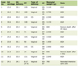
In clinical practice, numerous cases requiring differentiation between cutaneous and breast apocrine carcinoma in the axillary region have been reported. The reported cases are summarized in Table 1 [60–81]. We identified 22 English-language case reports of axillary apocrine carcinoma in a PubMed search through August 2026 using keywords related to “apocrine carcinoma” and “axilla,” regardless of the presumed origin [60–81]. Reports that did not describe the diagnostic process were excluded. Among the 23 patients, the median age was 61 (33–92) years, with 19 male (83%) and four female (17%) patients. The median tumor size was 40 (4–120) mm.
 Click to view | Table 1. Reported Cases of Axillary Apocrine Carcinoma |
Interestingly, in 21 (91%) of the 23 cases, both cutaneous and breast apocrine carcinoma were considered during the diagnostic process before one origin was ultimately favored. As the basis for diagnosis, 14 cases relied on the presence or absence of adjacent normal structures, including ectopic mammary tissue or normal apocrine glands. Seven cases cited the absence of a detectable breast primary lesion, either alone or in combination with pathological findings, as supportive evidence for diagnosis. In this differential diagnosis, three possibilities should be considered: primary cutaneous apocrine carcinoma, carcinoma arising in ectopic breast tissue, and metastatic breast carcinoma. The presence of adjacent cutaneous apocrine structures may support a primary cutaneous origin, whereas identification of ectopic mammary tissue or an in situ component favors carcinoma arising in ectopic breast tissue. In contrast, detection of a primary breast lesion supports metastatic breast carcinoma. On the other hand, several reported tumors exceeded 10 cm in diameter, raising the possibility that extensive tumor involvement may obscure or replace the surrounding normal structures. Therefore, absence of adjacent mammary tissue or apocrine glands should be interpreted cautiously when excluding a particular origin. Likewise, although absence of a detectable breast primary lesion may be useful in excluding metastatic breast carcinoma involving the skin or lymph nodes, this finding alone may not reliably distinguish cutaneous apocrine carcinoma from carcinoma arising in ectopic breast tissue. Although immunohistochemical findings such as GCDFP-15, cytokeratin 7 (CK7), and cytokeratin 20 (CK20) were reported in nearly all cases, only six reports explicitly described immunohistochemical findings as part of the basis for diagnosis.
| Immunohistochemical Overlap Between Cutaneous and Breast Apocrine Carcinoma and Practical Diagnostic Approach | ▴Top |
Because differentiation between cutaneous and breast apocrine carcinoma in the axillary region is often challenging based on histopathological findings alone, various immunohistochemical markers have been discussed in the literature as potential tools for determining tumor origin. Figure 3 summarizes previous studies involving cohorts of five or more cases of cutaneous and/or breast apocrine carcinoma that reported the positivity rates of immunohistochemical markers [4, 5, 82–103]. In this summary, markers with a reported positivity rate of ≥ 50% were visually categorized as “frequently positive” (shown in pink) whereas those with < 50% positivity were categorized as “less frequently positive” (shown in light blue). Because the reported cohort sizes varied substantially, this categorization should be interpreted as a visual overview rather than a strict diagnostic threshold. Also, several markers were evaluated only in one tumor type or in small cohorts, limiting direct comparison.
 Click for large image | Figure 3. Summary of published immunohistochemical marker positivity in cutaneous and breast apocrine carcinoma. Each cell represents the results from a single published study. The upper line indicates the number of positive cases divided by the total number of cases, and the lower line indicates the corresponding positivity rate (%) within each study. Marker names are shown in the center column. Positivity data for breast apocrine carcinoma are displayed on the left, whereas those for cutaneous apocrine carcinoma are displayed on the right. Markers with a reported positivity rate of ≥ 50% are shown in pink (frequently positive), whereas those with a positivity rate of < 50% are shown in light blue (less frequently positive). Superscript letters in the upper right corner of each cell correspond to the reference numbers listed below. Reference key: a[89]; b[86]; c[99]; d[101]; e[84]; f[4]; g[92]; h[83]; i[5]; j[93]; k[88]; l[97]; m[98]; n[91]; o[82]; p[87]; q[103]; r[95]; s[90]; t[96]; u[94]; v[102]. AR: androgen receptor; GCDFP-15: gross cystic disease fluid protein 15; ER: estrogen receptor; TRPS1: trichorhinophalangeal syndrome type 1; p63: tumor protein p63; CK5/6: cytokeratin 5/6; CK14: cytokeratin 14; AMACR: alpha-methylacyl-CoA racemase; PgR: progesterone receptor; HER2: human epidermal growth factor receptor 2; GATA3, GATA binding protein 3. |
Overall, previously reported immunohistochemical profiles show substantial overlap between cutaneous and breast apocrine carcinoma, particularly for commonly used apocrine markers such as GCDFP-15, GATA binding protein 3 (GATA3), AR, and mammaglobin. Many of the reported cases of axillary apocrine carcinoma in which differential diagnosis was discussed (Table 1) [60–81] also employed these markers during the diagnostic process. However, these markers may support apocrine differentiation but are insufficient to reliably determine the tissue of origin when used alone.
Apocrine markers
Several immunohistochemistry (IHC) markers are characteristically expressed in apocrine cells and apocrine tumors. Among them, AR plays a central role in apocrine biology. Because normal apocrine sweat glands differentiate during puberty under the influence of sex-related hormones such as androgens [12, 14], apocrine sweat glands characteristically express AR [15]. Accordingly, AR expression is frequently observed in cutaneous apocrine carcinoma [1]. Similarly, breast apocrine carcinoma acquires AR expression through apocrine metaplasia [16], and AR positivity is currently regarded as one of its defining immunophenotypic features [30].
Several downstream targets or associated proteins of AR signaling have likewise been linked to apocrine differentiation [93]. GCDFP-15, one of the most widely used apocrine markers in clinical practice, was originally identified in gross cyst fluid of the breast and was subsequently shown to be specifically expressed in apocrine metaplastic epithelium rather than normal mammary epithelium [104, 105]. Consistent with this biology, GCDFP-15 expression has been widely reported in breast apocrine carcinoma and normal cutaneous apocrine glands, leading to its recognition as a classical marker of apocrine cells [86, 92, 104]. Accordingly, frequent positivity for GCDFP-15 has been reported in both breast and cutaneous apocrine carcinoma.
Alpha-methylacyl-CoA racemase (AMACR), a mitochondrial and peroxisomal enzyme involved in fatty acid metabolism [92], was originally established as a diagnostic marker for AR-positive prostate carcinoma [106] and has subsequently been reported in AR-positive breast carcinomas with apocrine differentiation [107]. Recent studies have further demonstrated frequent AMACR expression in a broad spectrum of mammary apocrine lesions, including apocrine metaplasia, apocrine ductal carcinoma in situ, and invasive apocrine carcinoma [108]. Similarly, adipophilin, a lipid droplet–associated protein also expressed in sebaceous carcinoma [109], reflects the intracytoplasmic lipid accumulation characteristic of apocrine cells. Although adipophilin positivity in cutaneous apocrine carcinoma appears relatively limited [4, 110], higher expression has been reported in tumors with apocrine differentiation, including pulmonary carcinomas with apocrine features and breast apocrine carcinoma [90, 111].
Taken together, these markers are useful for characterizing apocrine tumors and distinguishing them from non-apocrine malignancies. However, because these proteins are broadly expressed in both normal apocrine glands and tumors with apocrine differentiation, their utility for determining tissue origin appears limited.
Hormone-related markers
In breast cancer, hormone receptor status and HER2 expression play central roles in clinicopathological classification and therapeutic decision-making. Historically, breast apocrine carcinoma has been defined using various criteria, including purely morphological definitions as well as definitions combining apocrine morphology with hormone receptor status [19]. In the current WHO classification, however, carcinoma with apocrine differentiation is characterized by apocrine morphology together with an ER-negative/AR-positive immunophenotype [30]. Accordingly, most studies have reported breast apocrine carcinoma to be predominantly ER-negative and progesterone receptor (PgR)-negative. In contrast, HER2 status appears more heterogeneous, with both HER2-positive and HER2-negative cases being reported.
In cutaneous apocrine carcinoma, more than 50% positivity for ER and PgR was reported in three and two of the five available studies, respectively. In comparative studies between breast and cutaneous apocrine carcinoma, ER negativity was more frequently observed in breast apocrine carcinoma than in cutaneous apocrine carcinoma. Studies evaluating HER2 expression in cutaneous apocrine carcinoma remain limited, and reported HER2 positivity rates have varied substantially, ranging from 0% [4] to 50% [95]. Interestingly, several case reports have described favorable responses of HER2-positive cutaneous apocrine carcinoma to anti-HER2 therapy using treatment strategies adopted from breast cancer [62, 112], suggesting potential therapeutic relevance of HER2 evaluation in these tumors.
Overall, however, the significance of ER, PgR, and HER2 expression for determining tissue origin remains unclear.
Glandular and skin-associated markers
In general, breast and cutaneous carcinomas are often distinguished using lineage-associated immunohistochemical markers [113, 114]. However, this distinction is less straightforward in apocrine carcinoma because both entities share glandular morphology and apocrine differentiation [89, 98]. GATA3, mammaglobin, and trichorhinophalangeal syndrome type 1 (TRPS1) are commonly used breast lineage-associated markers [115], but their expression in apocrine carcinoma is inconsistent. GATA3 and mammaglobin show variable positivity in both breast and cutaneous apocrine carcinoma. Although TRPS1 has been proposed as a relatively breast-specific marker [116] and was historically considered negative in cutaneous apocrine glands and cutaneous apocrine carcinoma [117], more recent studies have demonstrated TRPS1 expression in these lesions [118].
In contrast, tumor protein p63 (p63) [119, 120], cytokeratin 5/6 (CK5/6) [121], cytokeratin 14 (CK14) [98], and podoplanin [122] have been described as markers associated with cutaneous adnexal differentiation. These markers were more frequently positive in cutaneous apocrine carcinoma and generally negative in breast apocrine carcinoma. Although complete separation between the two entities was not achieved, these markers may still provide supportive evidence favoring cutaneous origin. On the other hand, epidermal growth factor receptor (EGFR), which has been regarded as a basal-associated marker, is expressed in normal apocrine sweat glands and has also been reported in cutaneous apocrine carcinomas, while relatively frequent expression has additionally been observed in mammary apocrine carcinoma [100, 123, 124]. Because the available studies were small and methodologically heterogeneous, these findings should be interpreted as general expression tendencies rather than strict diagnostic criteria.
Practical diagnostic approach
In practice, differentiation between cutaneous and breast apocrine carcinoma should be based on an integrated assessment rather than on any single finding. Clinical history, tumor location and morphology, the presence or absence of adjacent apocrine glands or ectopic mammary tissue, and immunohistochemical findings should all be considered together. Conventional imaging modalities, including mammography, ultrasonography, and magnetic resonance imaging (MRI), may be useful for identifying ectopic breast tissue [125] or detecting a primary breast lesion [126]. However, clinical features such as age, sex, and disease course are not necessarily specific to either tumor type, and substantial overlap also exists in morphological and immunohistochemical findings [4]. Therefore, definitive determination of the tissue of origin may remain challenging even after comprehensive evaluation.
| Conclusions and Future Directions | ▴Top |
Cutaneous and breast apocrine carcinomas are biologically distinct malignancies arising from different organs and exhibiting markedly different clinical behaviors, prognoses, and therapeutic strategies. However, these tumors show substantial overlap in histopathological appearance and immunophenotypic features because of their shared apocrine differentiation. Although both entities are rare, numerous reports have highlighted the diagnostic difficulty encountered in distinguishing them, particularly in the axillary region. While several immunohistochemical markers showed differences in expression patterns between the two entities, currently available evidence remains limited and insufficiently reliable for routine diagnostic use.
Recent advances in transcriptomic profiling may provide new opportunities to improve the differential diagnosis of rare tumors. In our recent study, we developed a transcriptome-based molecular differentiation score to distinguish cutaneous from breast apocrine carcinoma [127]. For rare tumors such as apocrine carcinoma, the limited availability of training datasets is often a major obstacle to developing transcriptome-based diagnostic models. To address this challenge, we constructed the differentiation score using large transcriptomic cohorts representing the entire spectrum of breast and cutaneous carcinomas and subsequently validated its performance in apocrine carcinoma. These findings suggest that transcriptome-based approaches can accurately distinguish cutaneous from breast apocrine carcinoma despite the scarcity of transcriptomic data. As transcriptomic profiling becomes increasingly incorporated into routine clinical practice, such molecular diagnostic approaches may become practical adjuncts to histopathological and immunohistochemical evaluation for distinguishing diagnostically challenging rare tumors, including apocrine carcinoma. Future large comparative studies and independent validation of molecular diagnostic tools, including transcriptome-based differentiation scores, will be important for establishing more objective and reliable diagnostic strategies for these challenging tumors.
Acknowledgments
None to declare.
Financial Disclosure
KT is supported by US National Institutes of Health grant numbers R01CA287731, R01CA250412, and R37CA248018, as well as US Department of Defense BCRP grants W81XWH-19-1-0674 and HT9425-24-1-0762. Roswell Park Comprehensive Cancer Center is supported by US National Institutes of Health Cancer Center Support Grant number P30CA016056.
Conflict of Interest
The authors declare no competing financial interests.
Author Contributions
KK conceptualized and designed the study, performed statistical and bioinformatic analyses, and wrote the original draft. KT provided supervision, contributed to study design, and critically revised the manuscript. RM, EE, MO, AY, KN, TK, IE, TI critically revised the manuscript. All authors approved the final manuscript.
Data Availability
The authors declare that data supporting the findings of this study are available within the article.
Abbreviations
AMACR: alpha-methylacyl-CoA racemase; AR: androgen receptor; CK5/6: cytokeratin 5/6; CK7: cytokeratin 7; CK14: cytokeratin 14; CK20: cytokeratin 20; ER: estrogen receptor; GATA3: GATA binding protein 3; GCDFP-15: gross cystic disease fluid protein 15; HER2: human epidermal growth factor receptor 2; IHC: immunohistochemistry; PDPN: podoplanin; PgR: progesterone receptor; SEER: Surveillance, Epidemiology, and End Results; TRPS1: trichorhinophalangeal syndrome type 1; WHO: World Health Organization
| References | ▴Top |
- Tsuruta S, Ogata D, Namikawa K, Nakano E, Yamazaki N. A review of cutaneous apocrine carcinoma: epidemiology, diagnosis, prognosis, and treatment options. Jpn J Clin Oncol. 2024;54(11):1135-1140.
doi pubmed - Cserni G. Histological type and typing of breast carcinomas and the WHO classification changes over time. Pathologica. 2020;112(1):25-41.
doi pubmed - Celis JE, Gromova I, Gromov P, Moreira JM, Cabezon T, Friis E, Rank F. Molecular pathology of breast apocrine carcinomas: a protein expression signature specific for benign apocrine metaplasia. FEBS Lett. 2006;580(12):2935-2944.
doi pubmed - Piris A, Peng Y, Boussahmain C, Essary LR, Gudewicz TM, Hoang MP. Cutaneous and mammary apocrine carcinomas have different immunoprofiles. Hum Pathol. 2014;45(2):320-326.
doi pubmed - Vranic S, Gatalica Z. An update on the molecular and clinical characteristics of apocrine carcinoma of the breast. Clin Breast Cancer. 2022;22(4):e576-e585.
doi pubmed - Chamberlain RS, Huber K, White JC, Travaglino-Parda R. Apocrine gland carcinoma of the axilla: review of the literature and recommendations for treatment. Am J Clin Oncol. 1999;22(2):131-135.
doi pubmed - Teixeira IP, Azzopardi C, Chowdhury R, Zsolt O, Castro M, Teh J. Ectopic breast tissue presenting as an enlarging abdominal mass. Radiol Case Rep. 2020;15(6):733-740.
doi pubmed - Hibbs RG. Electron microscopy of human apocrine sweat glands. J Invest Dermatol. 1962;38:77-84.
doi pubmed - Fakoya AO, Murphrey MB, Safadi AO, Vaidya T. Histology, Apocrine Gland. In: StatPearls. Treasure Island (FL): StatPearls Publishing, 2026. Available from: https://www.ncbi.nlm.nih.gov/books/NBK482199/.
- Sato K, Leidal R, Sato F. Morphology and development of an apoeccrine sweat gland in human axillae. Am J Physiol. 1987;252(1 Pt 2):R166-180.
doi pubmed - Hu Y, Converse C, Lyons MC, Hsu WH. Neural control of sweat secretion: a review. Br J Dermatol. 2018;178(6):1246-1256.
doi pubmed - Baker LB. Physiology of sweat gland function: The roles of sweating and sweat composition in human health. Temperature (Austin). 2019;6(3):211-259.
doi pubmed - Savic I, Berglund H, Gulyas B, Roland P. Smelling of odorous sex hormone-like compounds causes sex-differentiated hypothalamic activations in humans. Neuron. 2001;31(4):661-668.
doi pubmed - Wilke K, Martin A, Terstegen L, Biel SS. A short history of sweat gland biology. Int J Cosmet Sci. 2007;29(3):169-179.
doi pubmed - Beier K, Ginez I, Schaller H. Localization of steroid hormone receptors in the apocrine sweat glands of the human axilla. Histochem Cell Biol. 2005;123(1):61-65.
doi pubmed - Quinn CM, D'Arcy C, Wells C. Apocrine lesions of the breast. Virchows Arch. 2022;480(1):177-189.
doi pubmed - Alan LC. On the “pink” epithelium of the cystic breast and the staining of its granules. The Journal of Patholocy and Bacteriology. 1945;57:267-270.
doi - Durham JR, Fechner RE. The histologic spectrum of apocrine lesions of the breast. Am J Clin Pathol. 2000;113(suppl_1):S3-18.
doi pubmed - D'Arcy C, Quinn CM. Apocrine lesions of the breast: part 2 of a two-part review. Invasive apocrine carcinoma, the molecular apocrine signature and utility of immunohistochemistry in the diagnosis of apocrine lesions of the breast. J Clin Pathol. 2019;72(1):7-11.
doi pubmed - Wells CA, El-Ayat GA. Non-operative breast pathology: apocrine lesions. J Clin Pathol. 2007;60(12):1313-1320.
doi pubmed - Nielsen M, Thomsen JL, Primdahl S, Dyreborg U, Andersen JA. Breast cancer and atypia among young and middle-aged women: a study of 110 medicolegal autopsies. Br J Cancer. 1987;56(6):814-819.
doi pubmed - Toth J, Szamel I, Svastics E, DeSombre ER. Significance of apocrine metaplasia in mammary carcinogenesis. A preliminary morphological and immunohistochemical study. Ann N Y Acad Sci. 1990;586:238-251.
doi pubmed - Foote FW, Stewart FW. Comparative studies of cancerous versus noncancerous breasts. Ann Surg. 1945;121(2):197-222.
doi pubmed - Page DL, Dupont WD, Jensen RA. Papillary apocrine change of the breast: associations with atypical hyperplasia and risk of breast cancer. Cancer Epidemiol Biomarkers Prev. 1996;5(1):29-32.
pubmed - Fuehrer N, Hartmann L, Degnim A, Allers T, Vierkant R, Frost M, Visscher D. Atypical apocrine adenosis of the breast: long-term follow-up in 37 patients. Arch Pathol Lab Med. 2012;136(2):179-182.
doi pubmed - Carter DJ, Rosen PP. Atypical apocrine metaplasia in sclerosing lesions of the breast: a study of 51 patients. Mod Pathol. 1991;4(1):1-5.
pubmed - Calhoun BC, Booth CN. Atypical apocrine adenosis diagnosed on breast core biopsy: implications for management. Hum Pathol. 2014;45(10):2130-2135.
doi pubmed - Seidman JD, Ashton M, Lefkowitz M. Atypical apocrine adenosis of the breast: a clinicopathologic study of 37 patients with 8.7-year follow-up. Cancer. 1996;77(12):2529-2537.
doi pubmed - Plachta I, Kleibert M, Czarnecka AM, Spalek M, Szumera-Cieckiewicz A, Rutkowski P. Current diagnosis and treatment options for cutaneous adnexal neoplasms with apocrine and eccrine differentiation. Int J Mol Sci. 2021;22(10).
doi pubmed - WHO classification of tumours of the breast. 5 edn. International Agency for Research on Cancer. 2019.
- Gibbs DC, Yeung H, Blalock TW. Incidence and trends of cutaneous adnexal tumors in the United States in 2000-2018: A population-based study. J Am Acad Dermatol. 2023;88(1):226-228.
doi pubmed - Hollowell KL, Agle SC, Zervos EE, Fitzgerald TL. Cutaneous apocrine adenocarcinoma: defining epidemiology, outcomes, and optimal therapy for a rare neoplasm. J Surg Oncol. 2012;105(4):415-419.
doi pubmed - Ogata D, Kiyohara Y, Yoshikawa S, Kasami M. Treatment strategy for cutaneous apocrine carcinoma. Int J Clin Oncol. 2014;19(4):712-715.
doi pubmed - Tsuruta S, Ogata D, Namikawa K, Nakano E, Hiki K, Okumura M, Matsui Y, et al. Prognostic factors in cutaneous apocrine carcinoma: a single-institution retrospective study of 32 patients. Int J Clin Oncol. 2023;28(12):1690-1696.
doi pubmed - Fujisawa Y, Fujimoto M. Metastatic cutaneous apocrine carcinoma of the axilla successfully treated using systemic chemotherapy with i.v. epirubicin and cyclophosphamide followed by oral fluorinated pyrimidine. J Dermatol. 2014;41(3):280-282.
doi pubmed - Tlemcani K, Levine D, Smith RV, Brandwein-Gensler M, Staffenberg DA, Garg MK, Shifteh K, et al. Metastatic apocrine carcinoma of the scalp: prolonged response to systemic chemotherapy. J Clin Oncol. 2010;28(24):e412-414.
doi pubmed - Collette F, Hamoir M, Van Eeckhout P, D'Abadie P, Duprez T, Schmitz S, Machiels JP. Metastatic cutaneous apocrine adenocarcinoma successfully treated with systemic anti-androgen therapy-A case report. Clin Case Rep. 2020;8(12):3472-3478.
doi pubmed - Otsuka M, Yamasaki O, Kaji T, Shien T, Iwatsuki K. Metastatic cutaneous apocrine adenocarcinoma treated with a combination of pertuzumab-based targeted therapy and taxane chemotherapy: a case report. JAMA Dermatol. 2016;152(1):111-113.
doi pubmed - Frable WJ, Kay S. Carcinoma of the breast. Histologic and clinical features of apocrine tumors. Cancer. 1968;21(4):756-763.
doi pubmed - Arciero CA, Diehl AH, 3rd, Liu Y, Sun Q, Gillespie T, Li X, Subhedar P. Triple-negative apocrine carcinoma: a rare pathologic subtype with a better prognosis than other triple-negative breast cancers. J Surg Oncol. 2020;122(6):1232-1239.
doi pubmed - Saridakis A, Berger ER, Harigopal M, Park T, Horowitz N, Le Blanc J, Zanieski G, et al. Apocrine breast cancer: unique features of a predominantly triple-negative breast cancer. Ann Surg Oncol. 2021;28(10):5610-5616.
doi pubmed - Horimoto Y, Kumamaru H, Orimoto K, Ishikawa T, Kinukawa N, Iwamoto T, Taira N, et al. Chemotherapy is associated with differential survival outcomes in apocrine carcinoma: A nationwide cohort study from the Japanese National Clinical Database-Breast Cancer Registry. Breast. 2026;87:104779.
doi pubmed - Suzuki C, Yamada A, Kawashima K, Sasamoto M, Fujiwara Y, Adachi S, Oshi M, et al. Clinicopathological characteristics and prognosis of triple-negative apocrine carcinoma: a case-control study. World J Oncol. 2023;14(6):551-557.
doi pubmed - Wu W, Wu M, Peng G, Shi D, Zhang J. Prognosis in triple-negative apocrine carcinomas of the breast: a population-based study. Cancer Med. 2019;8(18):7523-7531.
doi pubmed - Kawashima K, Oshi M, Yamada A, Narui K, Shah Z, Khoury T, Endo I, et al. The clinical relevance of apocrine morphology in breast cancer depends on the estrogen receptor status. NPJ Breast Cancer. 2026;12(1).
doi pubmed - Hu T, Liu Y, Wu J, Hu XL, Zhao G, Liang B, Wang S, et al. Triple-negative apocrine breast carcinoma has better prognosis despite poor response to neoadjuvant chemotherapy. J Clin Med. 2022;11(6).
doi pubmed - Schwartz CJ, Ruiz J, Bean GR, Sirohi D, Joseph NM, Hosfield EM, Jacobs TW, et al. Triple-negative apocrine carcinomas: toward a unified group with shared molecular features and clinical behavior. Mod Pathol. 2023;36(5):100125.
doi pubmed - Hwang I, Lim Y, Song S, Lee H, Cho YA, Im YH, An JS, et al. Neoadjuvant chemotherapy response in triple-negative apocrine carcinoma: comparing apocrine morphology, androgen receptor, and immune phenotypes. Arch Pathol Lab Med. 2025;149(4):354-362.
doi pubmed - Cortes J, Rugo HS, Cescon DW, Im SA, Yusof MM, Gallardo C, Lipatov O, et al. Pembrolizumab plus chemotherapy in advanced triple-negative breast cancer. N Engl J Med. 2022;387(3):217-226.
doi pubmed - Takahashi N, Funasaka C, Shimura M, Hirota A, Uematsu M, Fukuda M, Baba K, et al. Efficacy of perioperative pembrolizumab for triple-negative breast cancer with apocrine feature or metaplastic carcinoma. Oncologist. 2026;31(6).
doi pubmed - Xiao X, Jin S, Zhangyang G, Xiao S, Na F, Yue J. Tumor-infiltrating lymphocytes status, programmed death-ligand 1 expression, and clinicopathological features of 41 cases of pure apocrine carcinoma of the breast: a retrospective study based on clinical pathological analysis and different immune statuses. Gland Surg. 2022;11(6):1037-1046.
doi pubmed - Groscurth P. Anatomy of sweat glands. Curr Probl Dermatol. 2002;30:1-9.
doi pubmed - Loukas M, Clarke P, Tubbs RS. Accessory breasts: a historical and current perspective. Am Surg. 2007;73(5):525-528.
pubmed - Ching D, Davies G. Primary ectopic breast carcinoma of the axilla in a male patient. J Surg Case Rep. 2017;2016(11).
doi pubmed - Fama F, Cicciu M, Sindoni A, Scarfo P, Pollicino A, Giacobbe G, Buccheri G, et al. Prevalence of ectopic breast tissue and tumor: a 20-year single center experience. Clin Breast Cancer. 2016;16(4):e107-112.
doi pubmed - Ehiagwina LA, Tagar E, Kpolugbo J, Oigbochie OE. Axillary breast: our limited experience of surgical excision. J Surg Case Rep. 2025;2025(7):rjaf536.
doi pubmed - Kurtzman JS, Pinkasovic E, Preminger BA. Treatment options for the clinical management of axillary breast tissue. Plast Reconstr Surg Glob Open. 2023;11(8):e5189.
doi pubmed - Marshall MB, Moynihan JJ, Frost A, Evans SR. Ectopic breast cancer: case report and literature review. Surg Oncol. 1994;3(5):295-304.
doi pubmed - Pang L, Cui M, Dai W, Wu S, Kong J. Diagnosis and treatment of male accessory breast cancer: a comprehensive systematic review. Front Oncol. 2021;11:640000.
doi pubmed - Mishima Y, Nakagawa T, Imai H, Sakurai F, Saito T. Accessory breast apocrine carcinoma: report of a rare case. Cureus. 2026;18(3):e104821.
doi pubmed - Kameh Khosh M, Hirsch T, Schulze HJ, Kuhnen C, Wellenbrock S. Case Report: Primary cutaneous apocrine carcinoma mimicking breast carcinoma - a rare diagnostic challenge. Front Oncol. 2025;15:1664122.
doi pubmed - Kamata A, Hino K, Matsuda A, Kamiyama K, Takasaka Y, Tazawa K. Treatment of HER2-positive cutaneous apocrine carcinoma of the axilla. Int Cancer Conf J. 2024;13(4):348-352.
doi pubmed - Khessairi N, Fertani Y, Sakhri S, Abbess I, Zemni I, Dhiab TB. Primary cutaneous apocrine carcinoma: a challenging case report. Int J Surg Case Rep. 2024;117:109513.
doi pubmed - Libertini M, Oneda E, Di Biasi B, Savelli G, Zaniboni A. Cutaneous adnexal carcinoma with apocrine differentiation: a challenging diagnosis and personalized treatment with mTOR inhibitor in a very rare disease. Case Rep Oncol. 2020;13(3):1091-1096.
doi pubmed - Sekine C, Kawase K, Yoshida K. Sentinel lymph node biopsy of primary apocrine sweat gland carcinoma of the axilla: a case report and review of the literature. Int J Surg Case Rep. 2020;77:122-125.
doi pubmed - Benzzi H, Askour M, Elouazani A, Bouaddi M, Hassam B, Saidi A. What do you think of an unusual axillary mass? Pan Afr Med J. 2017;28:309.
doi pubmed - Berdugo J, Dumont-Mackay V, Brissy-Lachery S, Leclerc YE, Doyon J, Mitchell A, Petrella T, et al. Cutaneous apocrine carcinoma with an in situ component and histiocytoid and signet-ring cells. Am J Dermatopathol. 2017;39(6):e76-e78.
doi pubmed - Seong MK, Kim EK, Han K, Seol H, Kim HA, Noh WC. Primary apocrine sweat gland carcinomas of the axilla: a report of two cases and a review of the literature. World J Surg Oncol. 2015;13:59.
doi pubmed - Kopecky J, Korena A, Priester P, Dolezal J, Kasparova P, Kyllarova A. Durable complete metabolic responses to androgen receptor-targeted therapy in metastatic primary cutaneous apocrine carcinoma: a case report and literature review. Discov Oncol. 2026;17(1).
doi pubmed - Li C, Qian B. Male triple negative axillary accessory breast cancer-a case report. Transl Cancer Res. 2022;11(8):2926-2930.
doi pubmed - Zahid R, Soofi ME, Elmalik H, Junejo K. Primary apocrine carcinoma of the axilla in a male patient: a case report. Clin Case Rep. 2016;4(4):344-347.
doi pubmed - Vasilakaki T, Skafida E, Moustou E, Grammatoglou X, Arkoumani E, Koulia K, Tsavari A, et al. Primary cutaneous apocrine carcinoma of sweat glands: a rare case report. Case Rep Oncol. 2011;4(3):597-601.
doi pubmed - Pucevich B, Catinchi-Jaime S, Ho J, Jukic DM. Invasive primary ductal apocrine adenocarcinoma of axilla: a case report with immunohistochemical profiling and a review of literature. Dermatol Online J. 2008;14(6):5.
pubmed - Gatti AP, Tonello L, Pfaffenzeller W, Savoia FO, Goergen DI, De Pieri Coan R, Teixeira UF, et al. Apocrine sweat gland adenocarcinoma: a rare case report and review. Int J Surg Case Rep. 2017;36:78-81.
doi pubmed - Ahmad A, Samat S, Tan Y, Bumpers H. Case Report: A challenging diagnosis of an apocrine sweat gland carcinoma. Front Surg. 2024;11:1307647.
doi pubmed - Lin Y, Wang Y. Case report of a male primary breast carcinoma of axillary accessory mammary gland. Clin Breast Cancer. 2012;12(2):142-144.
doi pubmed - Rina Fujiwara-Tani JH, Masayuki Ikeda, Kaori Hanaoka, Hiroki Kuniyasu. Axillary carcinoma with apocrine differentiation: a case report. Clinical and Diagnostic Pathology. 2018;2:1-3.
- Talukdar A, Khanna S, Purkayastha J, Das G, Ramchandani S. Axillary apocrine adenocarcinoma: a diagnostic challenge. Indian J Surg Oncol. 2022;13(2):356-358.
doi pubmed - Kathrotiya PR, Bridge AT, Warren SJ, Do H, Klenk AS, Xu LY, Mathur AN. Primary apocrine adenocarcinoma of the axilla. Cutis. 2015;95(5):271-274, 281.
pubmed - Toledo-Pastrana T, Llombart-Cussac B, Traves-Zapata V, Requena-Caballero C, Sanmartin-Jimenez O, Angeles-Sales M, Cabezas M, et al. Case report: differential diagnosis between primary cutaneous apocrine adenocarcinoma versus extramammary or metastatic breast adenocarcinoma. Am J Dermatopathol. 2014;36(10):e175-178.
doi pubmed - Zelger BG, Stelzmueller I, Dunst KM, Zelger B. Solid apocrine carcinoma of the skin: report of a rare adnexal neoplasm mimicking lobular breast carcinoma. J Cutan Pathol. 2008;35(3):332-336.
doi pubmed - Celis JE, Gromov P, Cabezon T, Moreira JM, Friis E, Jirstrom K, Llombart-Bosch A, et al. 15-prostaglandin dehydrogenase expression alone or in combination with ACSM1 defines a subgroup of the apocrine molecular subtype of breast carcinoma. Mol Cell Proteomics. 2008;7(10):1795-1809.
doi pubmed - Fernandez-Flores A. Immunohistochemical and morphologic evaluation of primary cutaneous apocrine carcinomas and cutaneous metastases from ductal breast carcinoma. Rom J Morphol Embryol. 2012;53(4):879-892.
pubmed - Fernandez-Flores A. Cutaneous metastases from breast carcinoma: calretinin expression and estrogen, progesterone and Her2/neu status of the metastases, compared to primary cutaneous apocrine tumors. Rom J Morphol Embryol. 2013;54(3 Suppl):695-699.
pubmed - Gromov P, Espinoza JA, Talman ML, Honma N, Kroman N, Timmermans Wielenga V, Moreira JM, et al. FABP7 and HMGCS2 are novel protein markers for apocrine differentiation categorizing apocrine carcinoma of the breast. PLoS One. 2014;9(11):e112024.
doi pubmed - Honma N, Takubo K, Akiyama F, Sawabe M, Arai T, Younes M, Kasumi F, et al. Expression of GCDFP-15 and AR decreases in larger or node-positive apocrine carcinomas of the breast. Histopathology. 2005;47(2):195-201.
doi pubmed - Ilhan B, Emiroglu S, Turkay R, Ilhan R. The role of histopathologic testing on apocrine carcinoma of the breast. Curr Probl Cancer. 2020;44(2):100501.
doi pubmed - Matsuo K, Fukutomi T, Hasegawa T, Akashi-Tanaka S, Nanasawa T, Tsuda H. Histological and immunohistochemical analysis of apocrine breast carcinoma. Breast Cancer. 2002;9(1):43-49.
doi pubmed - Mentrikoski MJ, Wick MR. Immunohistochemical distinction of primary sweat gland carcinoma and metastatic breast carcinoma: can it always be accomplished reliably? Am J Clin Pathol. 2015;143(3):430-436.
doi pubmed - Moritani S, Ichihara S, Hasegawa M, Endo T, Oiwa M, Shiraiwa M, Nishida C, et al. Intracytoplasmic lipid accumulation in apocrine carcinoma of the breast evaluated with adipophilin immunoreactivity: a possible link between apocrine carcinoma and lipid-rich carcinoma. Am J Surg Pathol. 2011;35(6):861-867.
doi pubmed - Moriya T, Sakamoto K, Sasano H, Kawanaka M, Sonoo H, Manabe T, Ito J. Immunohistochemical analysis of Ki-67, p53, p21, and p27 in benign and malignant apocrine lesions of the breast: its correlation to histologic findings in 43 cases. Mod Pathol. 2000;13(1):13-18.
doi pubmed - Nakamura H, Kukita Y, Kunimasa K, Kittaka N, Kusama H, Nakayama T, Tamaki Y, et al. alpha-Methylacyl-CoA racemase: a useful immunohistochemical marker of breast carcinoma with apocrine differentiation. Hum Pathol. 2021;116:39-48.
doi pubmed - Nishida H, Kato A, Kaimori R, Kawamura K, Daa T. Relationship between androgen receptor and androgen receptor-related protein expression in breast cancers focusing on morphologically identified carcinoma with apocrine differentiation. Sci Rep. 2025;15(1):2892.
doi pubmed - Paties C, Taccagni GL, Papotti M, Valente G, Zangrandi A, Aloi F. Apocrine carcinoma of the skin. A clinicopathologic, immunocytochemical, and ultrastructural study. Cancer. 1993;71(2):375-381.
doi pubmed - Plaza JA, Brenn T, Gru AA, Matoso A, Sheldon J, Sangueza M. Poorly differentiated cutaneous apocrine carcinomas: histopathological clues and immunohistochemical analysis for the diagnosis of this unusual neoplasm. Histopathology. 2023;82(2):276-284.
doi pubmed - Portelli F, Salvati L, Projetto E, Gori A, Scarfi F, Trane L, Lo Russo G, et al. A clinical, pathological and immunohistochemical series of 9 cases of primary cutaneous apocrine carcinomas of the head and neck. Australas J Dermatol. 2020;61(2):e189-e195.
doi pubmed - Robson A, Lazar AJ, Ben Nagi J, Hanby A, Grayson W, Feinmesser M, Granter SR, et al. Primary cutaneous apocrine carcinoma: a clinico-pathologic analysis of 24 cases. Am J Surg Pathol. 2008;32(5):682-690.
doi pubmed - Rollins-Raval M, Chivukula M, Tseng GC, Jukic D, Dabbs DJ. An immunohistochemical panel to differentiate metastatic breast carcinoma to skin from primary sweat gland carcinomas with a review of the literature. Arch Pathol Lab Med. 2011;135(8):975-983.
doi pubmed - Tsutsumi Y. Apocrine carcinoma as triple-negative breast cancer: novel definition of apocrine-type carcinoma as estrogen/progesterone receptor-negative and androgen receptor-positive invasive ductal carcinoma. Jpn J Clin Oncol. 2012;42(5):375-386.
doi pubmed - Vranic S, Marchio C, Castellano I, Botta C, Scalzo MS, Bender RP, Payan-Gomez C, et al. Immunohistochemical and molecular profiling of histologically defined apocrine carcinomas of the breast. Hum Pathol. 2015;46(9):1350-1359.
doi pubmed - Wang J, Peng Y, Sun H, Aung PP, Resetkova E, Yam C, Sahin AA, et al. TRPS1 and GATA3 expression in invasive breast carcinoma with apocrine differentiation. Arch Pathol Lab Med. 2024;148(2):200-205.
doi pubmed - Yoon EC, Wang G, Parkinson B, Huo L, Peng Y, Wang J, Salisbury T, et al. TRPS1, GATA3, and SOX10 expression in triple-negative breast carcinoma. Hum Pathol. 2022;125:97-107.
doi pubmed - Yu M, Miao X, Dong X, Wu K, Zhong G, Zhang M, Ma M, et al. Clinicopathological observation and initial exploration of characteristic biomarkers in breast apocrine carcinoma. J Transl Med. 2026;24(1).
doi pubmed - Haagensen DE, Jr., Dilley WG, Mazoujian G, Wells SA, Jr. Review of GCDFP-15. An apocrine marker protein. Ann N Y Acad Sci. 1990;586:161-173.
doi pubmed - Mazoujian G, Pinkus GS, Davis S, Haagensen DE, Jr. Immunohistochemistry of a gross cystic disease fluid protein (GCDFP-15) of the breast. A marker of apocrine epithelium and breast carcinomas with apocrine features. Am J Pathol. 1983;110(2):105-112.
pubmed - Lerner G, Tang H, Singh K, Golestani R, St Claire S, Humphrey PA, Lannin D, et al. AMACR expression is a potential diagnostic marker in apocrine lesions of breast, and is associated with high histologic grade and lymph node metastases in some invasive apocrine breast cancers. Clin Breast Cancer. 2023;23(2):199-210.
doi pubmed - Zhou M, Chinnaiyan AM, Kleer CG, Lucas PC, Rubin MA. Alpha-Methylacyl-CoA racemase: a novel tumor marker over-expressed in several human cancers and their precursor lesions. Am J Surg Pathol. 2002;26(7):926-931.
doi pubmed - Gatalica Z, Stafford P, Vranic S. Alpha-methylacyl-CoA racemase (AMACR) protein is upregulated in early proliferative lesions of the breast irrespective of apocrine differentiation. Hum Pathol. 2022;129:40-46.
doi pubmed - Ostler DA, Prieto VG, Reed JA, Deavers MT, Lazar AJ, Ivan D. Adipophilin expression in sebaceous tumors and other cutaneous lesions with clear cell histology: an immunohistochemical study of 117 cases. Mod Pathol. 2010;23(4):567-573.
doi pubmed - Boussahmain C, Mochel MC, Hoang MP. Perilipin and adipophilin expression in sebaceous carcinoma and mimics. Hum Pathol. 2013;44(9):1811-1816.
doi pubmed - Fujimoto M, Yoshizawa A, Sumiyoshi S, Sonobe M, Menju T, Hirata M, Momose M, et al. Adipophilin expression in lung adenocarcinoma is associated with apocrine-like features and poor clinical prognosis: an immunohistochemical study of 328 cases. Histopathology. 2017;70(2):232-241.
doi pubmed - Hidaka T, Fujimura T, Watabe A, Hashimoto A, Haga T, Onami K, Mizuashi M, et al. Successful treatment of HER-2-positive metastatic apocrine carcinoma of the skin with lapatinib and capecitabine. Acta Derm Venereol. 2012;92(6):654-655.
doi pubmed - Saeed S, Keehn CA, Morgan MB. Cutaneous metastasis: a clinical, pathological, and immunohistochemical appraisal. J Cutan Pathol. 2004;31(6):419-430.
doi pubmed - Prieto VG, Ivan D. The use of immunohistochemistry in the differential diagnosis of primary cutaneous adnexal neoplasms and metastatic adenocarcinomas to the skin. Diagnostic Histopathology. 2010;16:409-416.
doi - Yang SH, Lee JS, Koh JW, Nikas IP, Kim EN, Lee H, Ryu HS. Deciphering Breast Origin in Malignant Effusions: The Diagnostic Utility of an MGP, GATA-3, and TRPS-1 Immunocytochemical Panel. Pathobiology. 2025;92(1):40-51.
doi pubmed - Kong C, Yu B, Bi R, Xu X, Cheng Y, Yang W, Shui R. TRPS1, a sensitive marker for different histological and molecular types of breast cancer. Diagn Pathol. 2024;19(1):121.
doi pubmed - Abdelhammed MH, Cho WC. The evolving role of TRPS1 in dermatopathology: insights from the past 4 years. J Pathol Transl Med. 2026;60(2):129-143.
doi pubmed - Lenskaya V, Yang RK, Cho WC. Primary cutaneous apocrine carcinoma with RARA::NPEPPS fusion. J Cutan Pathol. 2024;51(6):419-423.
doi pubmed - Mahalingam M, Nguyen LP, Richards JE, Muzikansky A, Hoang MP. The diagnostic utility of immunohistochemistry in distinguishing primary skin adnexal carcinomas from metastatic adenocarcinoma to skin: an immunohistochemical reappraisal using cytokeratin 15, nestin, p63, D2-40, and calretinin. Mod Pathol. 2010;23(5):713-719.
doi pubmed - Lee JJ, Mochel MC, Piris A, Boussahmain C, Mahalingam M, Hoang MP. p40 exhibits better specificity than p63 in distinguishing primary skin adnexal carcinomas from cutaneous metastases. Hum Pathol. 2014;45(5):1078-1083.
doi pubmed - Plumb SJ, Argenyi ZB, Stone MS, De Young BR. Cytokeratin 5/6 immunostaining in cutaneous adnexal neoplasms and metastatic adenocarcinoma. Am J Dermatopathol. 2004;26(6):447-451.
doi pubmed - Liang H, Wu H, Giorgadze TA, Sariya D, Bellucci KS, Veerappan R, Liegl B, et al. Podoplanin is a highly sensitive and specific marker to distinguish primary skin adnexal carcinomas from adenocarcinomas metastatic to skin. Am J Surg Pathol. 2007;31(2):304-310.
doi pubmed - Saga K, Jimbow K. Immunohistochemical localization of activated EGF receptor in human eccrine and apocrine sweat glands. J Histochem Cytochem. 2001;49(5):597-602.
doi pubmed - Le LP, Dias-Santagata D, Pawlak AC, Cosper AK, Nguyen AT, Selim MA, Deng A, et al. Apocrine-eccrine carcinomas: molecular and immunohistochemical analyses. PLoS One. 2012;7(10):e47290.
doi pubmed - Salemis NS. Primary ectopic breast carcinoma in the axilla: A rare presentation and review of the literature. Breast Dis. 2021;40(2):109-114.
doi pubmed - de Bresser J, de Vos B, van der Ent F, Hulsewe K. Breast MRI in clinically and mammographically occult breast cancer presenting with an axillary metastasis: a systematic review. Eur J Surg Oncol. 2010;36(2):114-119.
doi pubmed - Kawashima K, Yamada A, Fallon EA, Fujita R, Sasamoto M, Oshi M, Narui K, et al. The development of Breast Apocrine Carcinoma (BAC) score that distinguishes breast and cutaneous apocrine carcinomas. World J Oncol. 2026, in press.
doi
This article is distributed under the terms of the Creative Commons Attribution 4.0 International License (CC BY 4.0), which permits unrestricted use, distribution, and reproduction in any medium, including commercial use, provided the original work is properly cited.
World Journal of Oncology is published by Elmer Press Inc.