| World Journal of Oncology, ISSN 1920-4531 print, 1920-454X online, Open Access |
| Article copyright, the authors; Journal compilation copyright, World J Oncol and Elmer Press Inc |
| Journal website https://wjon.elmerpub.com |
Original Article
Volume 17, Number 6, December 2026, pages 898-911

Dysregulated Serum miR-222-3p, miR-595, and miR-130a-3p as Potential Noninvasive Diagnostic Biomarkers for Papillary Thyroid Carcinoma
Shawnm Abdulah Ismaila, Parween Abdulsamad Ismailb, c
aDepartment of Nursing, Medical Technical Institute, Erbil Polytechnic University, Kurdistan Region, Iraq
bChemistry Department, College of Education, Salahaddin University-Erbil, Kurdistan Region, Iraq
cCorresponding Author: Parween Abdulsamad Ismail, Chemistry Department, College of Education, Salahaddin University-Erbil, Kurdistan Region, Iraq
Manuscript submitted July 31, 2026, accepted September 30, 2026, published online November 11, 2026
Short title: Serum miRNAs as Diagnostic Biomarkers for PTC
doi: https://doi.org/10.14740/wjon2858
| Abstract | ▴Top |
Background: Thyroid cancer is the most common malignancy in the endocrine system, with an annual incidence growing faster than nearly any other cancer. Papillary thyroid carcinoma (PTC) represents the most prevalent subtype of thyroid cancer. Accumulating evidence indicates that specific microRNAs (miRNAs) are consistently dysregulated in PTC. This study aimed to evaluate differentially expressed miRNAs in serum, which might become potential biomarkers for the diagnosis of PTC.
Methods: A total of 75 PTC patients attending the Smart Health Tower in Sulaymaniyah City and 55 healthy controls (HCs) from June 2024 and May 2025 were enrolled in this case-control study. Reverse transcription-quantitative polymerase chain reaction (RT-qPCR) was used to measure the serum levels of certain miRNAs (hsa-miR-222-3p, hsa-miR-595, and hsa-miR-130a-3p), with U6 serving as the internal reference. Using receiver operating characteristic (ROC) curve analysis, the diagnostic performance was determined.
Results: Three miRNAs presented significant dysregulation in PTC patients when compared with HC individuals (P < 0.0001). The miR-222-3p and miR-595 expression levels were upregulated with approximately 5-fold and 6.5-fold, respectively. Conversely, the miR-130a-3p expression level was downregulated by approximately 5-fold. ROC curve analysis demonstrated exceptional diagnostic value: miR-222-3p achieved an area under the curve (AUC) of 0.996 (95% confidence interval (CI), 0.994–0.999) with 98% sensitivity and 100% specificity; miR-595 showed an AUC of 0.970 (95% CI, 0.901–0.996) with 92% sensitivity and 96% specificity; and miR-130a-3p exhibited an AUC of 0.939 (95% CI: 0.600–0.900) with 92% sensitivity and 88% specificity. Similar expression patterns were identified across PTC, and the diagnostic performance remained consistent across all age and gender subgroups. Nonetheless, no statistically significant correlations between clinicopathological characteristics and miRNA expression were found.
Conclusions: Circulating serum levels of miR-222-3p, miR-595, and miR-130a-3p might serve as noninvasive diagnostic biomarkers for PTC. Elevated expression levels of miR-222-3p and miR-595 implicate an oncogenic role, whereas reduced levels of miR-130a-3p indicate a tumor-suppressive function in thyroid carcinogenesis. miRNAs could be essential molecular biomarkers for PTC diagnosis when applied in serum.
Keywords: hsa-miR-222-3p; hsa-miR-595; hsa-miR-130a-3p; Papillary thyroid carcinoma
| Introduction | ▴Top |
Thyroid carcinoma (TC) is one of the most common malignant neoplasms of the endocrine system. In 2020, an estimated that 586,000 new cases of TC were identified worldwide [1]. According to histological characteristics, TC can be divided into four primary groups: anaplastic thyroid carcinoma (ATC), medullary thyroid carcinoma (MTC), follicular thyroid carcinoma (FTC), and papillary thyroid carcinoma (PTC) [2]. Differentiated thyroid cancers (DTCs) include follicular and papillary thyroid cancers due to their similar features and clinical behavior, comprising almost 90% of all TC cases [3]. PTC accounts for most DTC diagnoses and is the most prevalent subtype of thyroid cancer, with its prevalence significantly rising in recent decades [4]. The increase in PTC diagnoses is primarily attributed to environmental influence and improvements in auxiliary diagnostic techniques [5]. Although the majority of DTC cases exhibit a favorable prognosis, particularly those with PTC, approximately 10% of patients diagnosed with PTC will exhibit metastatic progression or succumb to the disease during the course of cancer development [6]. Nonetheless, the outlook for most PTC patients is positive for long-term survival, despite a recurrence rate of 25–30% in PTC cases [7].
Ultrasound, frequently utilized in PTC screening, has identified thyroid nodules in up to 68% of assessed patients [4]. However, the gold standard for identifying thyroid nodules is fine-needle aspiration biopsy (FNAB); however, it has a number of drawbacks [8]. The size of the nodule, the anatomical location, and the cytopathologist’s level of experience can all affect the sensitivity and specificity of FNAB. Results might occasionally be inconclusive, necessitating additional testing or repeat biopsies. Moreover, it is a complex and invasive procedure associated with numerous potential complications [9, 10]. Notably, as many as 15–30% of the FNAB samples cannot accurately distinguish between benign and malignant lesions, resulting in several patients undergoing needless thyroidectomies for benign thyroid nodules [11]. Therefore, it is essential to identify sensitive and specific noninvasive diagnostic molecular biomarkers that can aid in the early diagnosis, prognosis, and monitoring of patients with PTC to prevent unnecessary surgeries and complications.
Endogenous, tiny, non-coding RNA molecules with a length of 19–25 nucleotides, microRNAs (also known as miRNAs or miRs) attach to the 3′ untranslated region (UTR) of target messenger RNAs (mRNAs) in the cytoplasm to act as post-transcriptional negative regulators of gene expression [12]. These miRNAs are involved in essential biological processes like tissue differentiation, metabolism, cellular life cycles, embryogenesis, and organogenesis. They exert critical regulatory functions on both oncogenes and tumor suppressor genes [13]. Recent results indicate that diverse molecular backgrounds, including dysregulated miRNA expressions, are prevalent across many diseases and cancer types [14], particularly in thyroid malignancies and their most prevalent histological subtype PTC [15]. Furthermore, the correlation of miRNAs with the pathogenesis of other thyroid cancer forms, including FTC, MTC, and ATC, has also been shown [13]. In cases when DTC was diagnosed, tumor progression could be attributed to a DICER1 RNase IIIb hotspot mutation that results in an imbalanced expression ratio of 5p:3p miRNAs [16].
PTC, distinguished by its unique molecular profile, frequently demonstrates changes in miRNA expression. The association between these miRNA alterations and disease progression is making specific miRNAs ideal candidates for being diagnostic or prognostic indicators [17, 18]. Previous studies of PTC tissue samples indicate that the expression levels of miR-146b, miR-221, miR-222, miR-375, miR-455-3p, and miR-551b-3p are significantly upregulated, whereas the expression levels of miR-204, miR-873, miR-532-5p, miR1296-5p, and miR-1301-3p are downregulated, potentially serving as biomarkers for diagnosing PTC patients [15, 19]. MiRNAs are considered to be highly stable molecules in tissues and biofluids (e.g., serum, plasma), with certain miRNAs being released from tissues into circulation with the disease progression, hence facilitating noninvasive biomarker identification [20]. Nonetheless, it is sometimes unfeasible to differentiate between benign and malignant tissue prior to surgery [4]. Conversely, blood samples are easily accessible, minimally invasive, and cost-effective, making them highly attractive for biomarker research [20, 21]. Moreover, a recent study by Verrienti et al (2025) investigated the potential of miR-146a-5p and miR-221 as novel diagnostic biomarkers for PTC using serum samples. The research indicated that these circulating miRNAs might accurately detect PTC and enhance the preoperative care of patients [22]. However, there are currently limited investigations of circulating miRNAs for the diagnosis of DTC (PTC and FTC), and the findings are inconsistent.
In this study, we focused on three specific miRNAs (miR-222-3p, miR-595, and miR-130a-3p). The rationale for their selection was twofold: first, our initial bioinformatic analysis of public expression repositories highlighted them as top-tier candidates; second, emerging literature has functionally implicated them in cell proliferation, apoptosis, invasion, and metastasis. By combining bioinformatic priority with verified literature-based relevance, we aimed to test their clinical applicability as circulating diagnostic tools.
This study sought to determine the biomarkers for the early detection of PTC as novel, noninvasive diagnostics by analyzing the expression levels and clinical significance of three miRNA molecules (hsa-miR-222-3p, hsa-miR-595, and hsa-miR-130a-3p) in serum samples and assessing the differences between patients with PTC and healthy controls (HCs).
| Materials and Methods | ▴Top |
Patient studies and clinical samples
This study was designed as a case-control investigation comprising a total of 130 participants. The study was conducted at the Smart Health Tower, Sulaymaniyah City, between June 2024 and May 2025. The patient group consisted of 75 individuals (50 females and 25 males) with a histopathologically confirmed diagnosis of PTC. An additional 55 age- and gender-matched HCs (35 females and 20 males) were included for comparative analysis. The HC group comprised individuals with no history of thyroid disease or malignancy, and no family history of thyroid disorders. HCs were recruited from the health examination center at the same institution during the same study period. Eligible controls had: (1) normal thyroid ultrasound findings showing no thyroid nodules; (2) normal serum thyroid hormone profiles (thyroid-stimulating hormone (TSH), free triiodothyronine (FT3), free thyroxine (FT4)); and (3) no history of thyroid disease or family history of thyroid dysfunction. Inclusion criteria were: (1) age >18 years; (2) primary diagnosis of PTC; and (3) preoperative blood sample collection. Patients were excluded if they presented with: (1) non-PTC thyroid malignancies; (2) secondary or recurrent PTC; (3) a prior history of neoadjuvant therapy, radiotherapy, or chemotherapy; (4) autoimmune conditions; or (5) concurrent secondary malignancies. The definitive reference standard for PTC diagnosis was postoperative histopathological confirmation evaluated by two experienced pathologists, classified in accordance with the World Health Organization (WHO) diagnostic criteria. All clinical staging was performed based on the eighth edition of the American Joint Committee on Cancer (AJCC)/tumor-node-metastasis (TNM) staging system.
Serum samples from PTC patients were collected 1 day before total thyroidectomy. Demographic data (age, gender) and primary tumor histology (tumor size, lymph node metastasis, TNM stage) were retrieved from patient medical records. The eighth edition of the AJCC staging manual was used to categorize the cancer stages [23]. All subjects provided written informed consent, and the study methodology was approved by the local Ethics Committee Salahaddin University (Approval No. 21008/2024). The study was conducted in accordance with the ethical standards of the Salahaddin University committee and the principles of the Declaration of Helsinki and its subsequent amendments. Blood samples (5 mL) were obtained from all participants, and the serum was isolated following centrifugation at 3,000 rpm for 10 min at 4 °C. An aliquot of 200 µL of fresh serum was immediately used for miRNA extraction and cDNA synthesis, while the remaining sera were stored in aliquots at −80 °C for further analysis. To control for the confounding effects of pre-analytical hemolysis on circulating miRNA levels, all plasma/serum samples underwent strict quality control screening. Hemolysis was assessed by visual inspection. Samples showing signs of hemolysis were excluded from subsequent RNA extraction and miRNA profiling.
The sample size was calculated using G*Power software (v3.1). An independent-samples t-test with a two-tailed alpha of 0.05 and a total sample size of 130 achieved 80.4% statistical power.
Total RNA extraction
Total RNA was isolated from serum samples with the Add Prep Total RNA Extraction Kit (Product Code: 10119, Korea). In brief, 200 µL of serum was mixed with 400 µL of lysis buffer, and the pellet was subsequently resuspended by vortexing. Then, 4 µL of β-mercaptoethanol and 20 µL of proteinase K (20 mg/mL) were added at 56 °C for 10 min to complete the dissociation of nucleoprotein complexes. Further processing of the samples was performed according to the manufacturer’s protocol. The isolated RNA was eluted in 50 µL of RNase-free water. The concentration and purity of the extracted RNA were assessed using the NanoDrop ND-1000 spectrophotometer (Biometrics, Wilmington, USA) by measuring the optical density at 260 nm and 280 nm (A260/280 ratio). After the extraction, the samples were promptly processed or stored at −80 °C.
Reverse transcription-quantitative polymerase chain reaction (RT-qPCR)
Complementary deoxyribonucleic acid (cDNA) was synthesized from the extracted RNA utilizing the AddScript Kit (Product Code: 22701, Korea) in accordance with the manufacturer’s guidelines. For each reaction, cDNA synthesis was performed as follows: 10 µL of 2 × reaction buffer, 2.0 µL of 10 mM dNTPs, 2.0 µL of specific stem-loop primers, 4 µL of RNA template, and 1.0 µL of 20 × AddScript enzyme solution, with 1 µL of nuclease-free water in a total volume of 20 µL. The miRNA primer sequences that were used in the study are presented in Table 1. Reverse transcription (RT) reaction was carried out in a thermal cycler with the following steps: priming at 25 °C for 10 min, RT at 50 °C for 60 min, RT enzyme inactivation at 80 °C for 5 min, and holding at 12 °C indefinitely. The generated cDNA was stored at −20 °C until analysis.
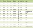 Click to view | Table 1. Oligonucleotide Primers Used in RT-qPCR for microRNA Expression |
Quantitative real-time PCR (qPCR) reaction was conducted in a final volume of 25 µL using RealQ Plus 2 × Master Mix SYBR Green (Amplicon, Denmark) following the manufacturer’s protocol. Each reaction mixture comprised 12.5 µL of PCR master mix, 1 µL of each primer, 3 µL of cDNA as a template, and 7.5 µL of PCR-grade water. A negative control containing all reagents except target miRNA was included to detect contamination in each qPCR assay. The qPCR was evaluated using the model version (Biorad CFX96 Real-time PCR System, USA), and all the reaction conditions were set up as follows: 95 °C for 3.5 min, followed by 40 cycles of denaturation at 95 °C for 38 s, annealing temperature for 30 s (61 °C for hsa-miR-595, 62 °C for hsa-miR-222-3p, 57 °C for hsa-miR-130a-3p, and 59 °C for U6), and 40 s at 72 °C with fluorescence readings during extension. All analyses were performed in duplicate to remove any outliers. Melting curve analysis (MCA) was performed from 65 °C to 95 °C with 0.5 °C increments every 5 s to evaluate the specificity of the PCR products. The cycle threshold (Ct) values for all samples were normalized to U6 as the endogenous control, and the expression levels of miRNAs were determined using the comparative 2−(ΔΔCt) method (ΔCt = CtmiRNA − CtU6).
Data analysis and fold change calculation
Relative miRNA expression levels were analyzed using the comparative cycle threshold (ΔΔCt) method, as described by Livak and Schmittgen (2001). The cycle threshold (Ct) represents the PCR cycle number at which the fluorescence signal rises above the background level. The ΔCt value was calculated by subtracting the Ct value of the endogenous reference gene RNU6-1 (U6) from the Ct value of each target miRNA (Livak and Schmittgen, 2001).
A fold change greater than 1 was considered indicative of upregulation, whereas a fold change less than 1 was indicative of downregulation of the target miRNA relative to the untreated control group.
Statistical analysis
All statistical analyses were performed using GraphPad Prism (version 10.4.2, GraphPad Software, USA) and MedCalc Statistical Software (version 20.215, MedCalc Software, Belgium). Relative expression levels of hsa-miR-222-3p, hsa-miR-595, and hsa-miR-130a-3p were quantified using the comparative 2−ΔΔCt method, with RNU6-1 (U6 small nuclear RNA) serving as the endogenous reference gene to normalize for inter-sample variation in cDNA input. Before inferential analyses, the normality of all continuous variables was assessed using the Shapiro–Wilk test. Because the data did not meet the assumption of normality, non-parametric statistical methods were applied throughout. Differences in miRNA expression levels, thyroid function parameters, and clinicopathological features between PTC patients and HCs were evaluated using the Mann–Whitney U test, whilst comparisons across multiple independent subgroups were conducted using the Kruskal–Wallis test. Categorical and demographic variables were compared using Fisher’s exact test. Continuous data are presented as mean ± standard error of the mean (SEM), whereas non-normally distributed data are expressed as median with interquartile range (IQR). The diagnostic performance of each miRNA was evaluated by receiver operating characteristic (ROC) curve analysis, from which the area under the curve (AUC) with 95% confidence interval (CI), sensitivity, specificity, and optimal cut-off values were determined.
| Results | ▴Top |
Demographic characteristics of patients with PTC and control group
Table 2 presents the baseline demographic and clinical characteristics of the study participants, comparing 75 patients with 55 controls. The variables assessed include age, gender, smoking status, alcohol consumption, physical activity, and family history of thyroid cancer. Overall, most characteristics were comparable between the two groups, except for family history of TC, which showed a statistically significant difference. The majority of participants in both groups were aged ≤ 45 years. Among patients, 50 (66.67%) were ≤ 45 years compared with 35 (63.64%) controls, while 25 (33.33%) patients and 20 (36.36%) controls were > 45 years. The difference was not statistically significant (P = 0.5018), indicating that age distribution was similar between patients and controls. This suggests that age was unlikely to act as a confounding factor in this study.
Click to view | Table 2. Baseline Demographic and Clinical Characteristics of Study Participants |
Females predominated in both groups, particularly among patients (66.67%) compared with controls (63.64%), while males accounted for 33.33% of patients and 36.36% of controls. However, this difference was not statistically significant (P = 0.5109). The higher proportion of females aligns with the known epidemiology of thyroid cancer, which occurs more frequently in women. Most participants in both groups were non-smokers. Among patients, 57 (76.00%) were non-smokers compared with 42 (76.36%) controls. Smokers comprised 18 (24.00%) of patients and 13 (23.64%) of controls. There was no statistically significant difference (P = 0.642), suggesting that smoking status was comparable between the groups.
Evaluation of miRNA relative expression in all PTC and HC
The expression levels of three miRNAs revealed statistically significant dysregulation in PTC patients compared with the HCs (P < 0.0001 for all comparisons), as summarized in Table 3. Specifically, hsa-miR-222-3p demonstrated substantial upregulation in the PTC group (5.275 ± 4.359) compared with healthy individuals (1.041 ± 0.335), representing an approximately 5-fold increase in expression levels (Fig. 1a). Similarly, hsa-miR-595 showed marked overexpression in the patient population (6.68 ± 2.752) relative to controls (1.061 ± 0.584), with nearly 6.5-fold elevated expression (Fig. 1b). Conversely, hsa-miR-130a-3p exhibited a distinct expression pattern, showing significant downregulation in PTC patients (0.205 ± 0.1445) compared with the HC (1.178 ± 0.9345), indicating an approximately 5-fold reduction in expression (Fig. 1c). These findings indicate that hsa-miR-222-3p and hsa-miR-595 may function as oncogenic miRNAs or disease-promoting factors, while hsa-miR-130a-3p appears to function as a tumor suppressor miRNA or protective factor.
Click to view | Table 3. The expression Profiles of the Three microRNAs in PTC Patients Compared With the HC |
 Click for large image | Figure 1. The figure demonstrates the comparison of relative expression (2−ΔΔCT values) in PTC patients and the healthy group. (a) hsa-miR-222-3p demonstrated substantial upregulation in the PTC group (5.501 ± 2.331) compared with healthy individuals (1.095 ± 0.3532), representing an approximately 5-fold increase in expression levels. (b) hsa-miR-595 showed marked overexpression in the patient population (6.876 ± 1.635) relative to controls (1.062 ± 0.3477), with nearly 6.5-fold elevated expression. (c) hsa-miR-130a-3p exhibited a distinct expression pattern, showing significant downregulation in PTC patients (0.2343 ± 0.1601) compared with the HC (1.241 ± 0.6993), indicating an approximately 5-fold reduction in expression. HC: healthy control; PTC: papillary thyroid carcinoma. |
The diagnostic value of the three miRNAs in PTC patients
ROC curve analysis was performed to evaluate the diagnostic efficacy of three circulating miRNAs, as illustrated in Table 4. The analysis examined that hsa-miR-222 3p exhibited exceptional discriminatory ability with AUC of 0.996 (95% CI, 0.994–0.999; P < 0.001), achieving near-perfect diagnostic value with 98% sensitivity and 100% specificity at an optimal cut-off value of > 1.769. The hsa-miR-595 showed a slightly lower AUC of 0.970 (95% CI, 0.901–0.996; P < 0.001), demonstrating 92% sensitivity and 96% specificity at a cut-off threshold of > 1.533, indicating excellent diagnostic ability but being slightly less precise than hsa-miR-222-3p. Additionally, hsa-miR-130a-3p demonstrated a strong diagnostic value with a moderate AUC of 0.939 (95% CI, 0.600–0.900; P < 0.001) and maintained 92% sensitivity with 88% specificity at a cut-off value of ≤ 0.425, using a downregulation threshold rather than upregulation. All three ROC curves were substantially above the diagonal reference line, and the statistically significant P values < 0.001 confirmed the robust discriminatory power of these miRNAs in diagnosing PTC patients very well from HC, as shown in Figure 2. As a whole, the results suggested that hsa-miR-222-3p, hsa-miR-595, and hsa-miR-130a-3p were effective noninvasive diagnostic biomarkers for PTC; among them, hsa-miR-222-3p showed the highest performance as a biomarker.
Click to view | Table 4. The analysis of ROC Curves for the Diagnostic Value of Three Selected miRNAs Markers in PTC |
 Click for large image | Figure 2. Receiver operating characteristic (ROC) curve analysis was conducted to assess the diagnostic performance of hsa-miR-222-3p, hsa-miR-595, and hsa-miR-130a-3p. The analysis evaluated that hsa-miR-222 3p exhibited exceptional discriminatory ability with an area under the curve (AUC) of 0.996 (95% CI, 0.994–0.999; P < 0.001), achieving near-perfect diagnostic value with 98% sensitivity and 100% specificity at an optimal cut-off value of > 1.769. The hsa-miR-595 showed a slightly lower AUC of 0.970 (95% CI, 0.901–0.996; P < 0.001), demonstrating 92% sensitivity and 96% specificity at the cut-off threshold of > 1.533, indicating excellent diagnostic ability but was slightly less precise than hsa-miR-222-3p. Additionally, hsa-miR-130a-3p demonstrated a strong diagnostic value with a moderate AUC of 0.939 (95% CI, 0.600–0.900; P < 0.001) and maintained 92% sensitivity with 88% specificity at a cut-off value of ≤ 0.425, using a downregulation threshold rather than upregulation. CI: confidence interval. |
Combined panel of miRNA as a diagnostic biomarker of PTC
Figure 3 presents the ROC analysis of the combined miRNAs for the diagnosis of PTC, evaluating their diagnostic performance in terms of the AUC, sensitivity, and specificity. The combined analysis of the three miRNAs (hsa-miR-222-3p, hsa-miR-595, and hsa-miR-130a-3p) demonstrated excellent diagnostic performance, with an AUC of 0.996, a sensitivity of 85.7%, and a specificity of 100%. These findings suggest that combining the three miRNAs may improve diagnostic specificity compared with individual miRNAs.
 Click for large image | Figure 3. ROC curve for PTC patients of combined hsa-miR-222-3p, hsa-miR-595, and hsa-miR-130a-3p. The combined analysis of all miRNAs significantly enhanced the diagnostic performance, achieving the highest AUC of 0.968, sensitivity of 0.857, and specificity of 1.0. AUC: area under the curve; ROC: receiver operating characteristic; PTC: papillary thyroid carcinoma. |
Evaluation of miRNA expression in all PTC versus HC based on demographic subgroups
The comparative analysis of miRNA expression levels (miR-222, miR-595, and miR-130a-3p) between PTC patients and HC was performed across different age and sex categories, with P values indicating statistical significance as shown in Table 5. In the participants aged ≤ 45 years (n = 50 patients, n = 35 controls) (Fig. 4a–c), as well as those > 45 years (n = 25 patients, n = 20 controls), remarkably similar relative expression was demonstrated (Fig. 4d–f). Specifically, miR-222-3p was shown to be significantly upregulated in the PTC patients, with average fold changes of 5.09 ± 4.295 and 5.459 ± 4.849 for younger and older groups, respectively, relative to the control group values of approximately 1.0 (P < 0.0001 for both comparisons). Similarly, miR-595 expression pattern was found to be elevated in patients across both age strata, with expression levels reaching 6.734 ± 2.785 and 6.413 ± 3.106 in the respective patient versus 1.103 ± 0.5735 and 1.061 ± 0.7875 in HCs (P < 0.0001). In contrast, miR-130a-3p revealed marked downregulation in the patient group, with mean values of 0.198 ± 0.144 and 0.221 ± 0.182, compared with values exceeding 1.0 in HCs (P < 0.0001). Sex-stratified analysis confirmed these findings, with male (n = 25 patients, n = 20 controls) and female (n = 50 patients, n = 35 controls) subgroups exhibiting nearly identical expression profiles of three selected miRNAs, as shown in Figure 5a–f, suggesting that the observed dysregulation is independent of demographic features.
Click to view | Table 5. The Comparative Expression of hsa-miR-222-3p, hsa-miR-595, and hsa-miR-130a-3p in PTC Patients and HC According to Age and Gender |
 Click for large image | Figure 4. Differential expression profiles of circulating miRNAs in PTC (n = 75) compared with HC samples (n = 55) stratified by age. (a) miR-222-3p age ≤ 45 years. (b) miR-595 age ≤ 45 years. (c) miR-130a-3p age ≤ 45 years. (d) miR-222-3p age > 45 years. (e) miR-595 age > 45 years. (f) miR-130a-3p age > 45 years. miR-222-3p was shown to be significantly upregulated in the DTC, with the average fold change of 5.408 ± 2.296 and 5.718 ± 2.476 for younger and older groups, respectively, relative to the control group values of approximately 1.0 (P < 0.0001 for both comparisons). miR-595 expression pattern was found to be elevated in patients across both age strata, with expression levels reaching 6.897 ± 1.62 and 6.827 ± 1.726 in the respective patient versus 1.1 ± 0.3339 and 0.9936 ± 0.3814 in healthy controls (P < 0.0001). miR-130a-3p revealed marked downregulation in the patient group, with mean values of 0.2267 ± 0.1434 and 0.2521 ± 0.1983 compared with HC exceeding 1.0 (P < 0.0001). miRNA: microRNA; PTC: papillary thyroid carcinoma; DTC: differentiated thyroid cancers; HC: healthy control. |
 Click for large image | Figure 5. Gender stratified differential relative expressions of three miRNAs in PTC patients compared with the HCs. (a) miR-222-3p in males. (b) miR-222-3p in females. (c) miR-595 in males. (d) miR-595 in females. (e) miR-130a-3p in males. (f) miR-130a-3p in females. Data are presented as mean ± SEM; P value < 0.0001. Sex-stratified analysis confirmed these findings, with male (n = 25 patients, n = 20 controls) and female (n = 50 patients, n = 35 controls) subgroups exhibiting nearly identical expression profiles of three selected miRNAs. HC: healthy control; miRNA: microRNA; PTC: papillary thyroid carcinoma; SEM: standard error of the mean. |
Clinicopathological characteristics of PTC and relative expression of hsa-miR-222-3p, hsa-miR-595, and hsa-miR-130a-3p in serum samples
Table 6 summarizes the association between clinicopathological characteristics of PTC and the serum expression levels of hsa-miR-222-3p, hsa-miR-595, and hsa-miR-130a-3p. Expression levels were calculated using the 2−ΔΔCT method and presented as mean ± SEM. Statistical significance was set at P < 0.05. Patients were grouped into micro/small tumors (≤ 2 cm, n = 28) and larger tumors (> 2 cm, n = 47). Expression differences between groups were modest. Although miR-595 showed slightly lower expression in larger tumors, the difference was not statistically significant. These miRNAs do not appear to correlate with tumor size, suggesting they may not reflect tumor burden. Patients were stratified into: N0 (no metastasis), N1a (central compartment) and N1b (lateral compartment). All P values exceeded 0.05. Although minor fluctuations were observed among groups, there was no statistically significant association between miRNA expression and lymph node status. Tumors were staged according to the AJCC TNM system. Although stage IV showed a numerically higher mean for miR-130a-3p, variability was high and statistical significance was not reached. Serum expression levels of these miRNAs are not significantly associated with tumor stage. Therefore, they do not appear to reflect disease progression severity in this cohort.
Click to view | Table 6. Clinicopathological Characteristics of PTC and Relative Expression of hsa-miR-222-3p, hsa-miR-595, and hsa-miR-130a-3p in Serum Samples |
Although these miRNAs may still have diagnostic potential (e.g., distinguishing PTC patients from HCs), this table suggests they lack prognostic value for stratifying patients by clinicopathological severity. The absence of correlation with tumor burden or metastatic status may indicate that these miRNAs are involved in early tumorigenesis rather than progression, or that their serum levels are influenced by systemic rather than tumor-specific mechanisms. The relatively small subgroup sizes (e.g., stage I and IV, N0 and N1b) may limit statistical power. Larger, multicenter cohorts may be required to detect subtle associations. Table 6 demonstrates that serum levels of hsa-miR-222-3p, hsa-miR-595, and hsa-miR-130a-3p are not significantly associated with clinicopathological characteristics of PTC. While these miRNAs may serve as potential biomarkers for PTC presence, they do not appear to reflect disease severity, metastatic status, or stage within this patient population.
| Discussion | ▴Top |
This study analyzed the relative expression of three miRs (miR-222-3p, miR-595, and miR-130a-3p) in serum samples of PTC patients compared with HC group, showing significant dysregulation patterns that underscore their potential as diagnostic biomarkers in PTC. The observed upregulation of miR-222-3p by approximately 5-fold in PTC patients exhibited exceptional diagnostic performance, with an AUC of 0.996, a sensitivity of 98%, and specificity of 100%. This finding is corroborated by recent meta-analytical evidence proving a high diagnostic value of the miR-221/222 cluster in PTC across multiple populations, suggesting the specificity could reach 100% and the sensitivity of 94.3% [24]. In addition, the overexpression of miR-222-3p validates findings of Armos et al (2024), who identified miR-222-3p among 30 significantly dysregulated miRNAs in 118 matched PTC tissues, establishing its consistent aberrant expression in TC and supporting its potential as a diagnostic marker [15].
The upregulation of miR-222-3p aligns with previous studies identifying it as a key oncogenic factor in TC, facilitating radioresistance and modulates critical signaling pathways including SOCS3/STAT3 [25]. miR-222-3p has demonstrated to promote proliferation, invasion, and migration of PTC by downregulating PTEN expression and activating the AKT signaling pathway [26, 27]. Supporting these mechanistic investigations, Matos et al (2024) found that miR-222-3p was significantly overexpressed in both cytological and histological samples of DTC with 73% sensitivity, and strongly associated with extrathyroidal extension, vascular invasion, and BRAF mutations [28]. A similar study by Kazlauskiene et al (2024) revealed that PTC patients had significantly higher plasma levels of miR-221-5p and miR-222-3p than HC, establishing them as novel markers for early diagnosis and monitoring of TC [29]. Moreover, the observed upregulation of miR-222-3p in our PTC patient cohort is consistent with a recent study demonstrating that serum miR-221-3p, a closely related member of the miR-221/222 cluster, efficiently discriminates benign from malignant thyroid nodules, presenting a novel diagnostic biomarker for PTC [22].
Beyond PTC, limited but significant information is available on the role of miR-222 in FTC. Nikiforova et al (2008) discovered that miR-221 and miR-222 were elevated in FTC [30]. However, Jikuzono et al (2013) documented substantial overexpression of miR-222 in metastatic FTC, whereby this miRNA facilitates carcinogenesis by targeting CDKN1B/CDKN1C and influencing metalloproteinase pathways [31]. Therefore, these miRNAs seem to be directly linked to the pathophysiology of both PTC and FTC, indicating wider diagnostic relevance for TC.
Similarly, the upregulation of miR-595 in PTC patients, exhibiting an approximate 6.5-fold increase relative to HC groups, represents a novel finding in TC research. Although limited literature exists specifically investigating miR-595 in thyroid cancers, its expression is elevated in PTC tissue samples, where it promotes cell migration by downregulating the sex-determining region Y-box 17 (Sox17) and consequently causing overexpression of the inflammatory cytokine interleukin-22 [32]. Krissansen et al (2015) examined the expression of miR-595 and found it to be significantly elevated in sera of patients with active inflammatory bowel disease compared with controls, where it targets NCAM-1 and FGFR2 to disrupt epithelial barrier integrity and promote disease pathogenesis [33]. Furthermore, Hao et al (2016) showed that miR-595 expression was upregulated in glioblastoma cells and tissues. The overexpression of miR-595 ameliorated glioblastoma cell proliferation and colony formation by suppressing SOX7 [34]. The strong diagnostic efficacy observed in our cohort (AUC = 0.970, sensitivity = 92%, specificity = 96%) aligns with results in hepatocellular carcinoma (HCC), where miR-595 attained an AUC of 0.92 in differentiating cirrhotic patients with HCC from those without malignancy, surpassing conventional biomarkers [35].
In contrast, miR-595 showed tumor suppressor properties, as it was found to be downregulated in HCC tissue and associated with poor overall survival, with studies showing that miR-595 inhibits cell proliferation, migration and invasion through suppressing of the NF-κB signaling pathway by targeting ABCB1 [36]. Similar results were described by Tian et al (2016), who reported that the expression of miR-595 was significantly lower in epithelial ovarian cancer tissue compared with noncancerous ovarian tissues [37]. These findings suggested that miR-595 showed context-dependent roles in different cancer types, functioning as an oncogene in TC while exhibiting tumor suppressor activity in hepatocellular and ovarian cancers, warranting further validation in larger multicenter studies.
Conversely, the substantial downregulation of miR-130a-3p in PTC patients, showing approximately a fivefold reduction compared with HC, suggests that this miRNA may function as a potential tumor suppressor in thyroid cancers. miR-130a-3p was significantly downregulated in PTC tissue compared with adjacent normal tissue, indicating a tumor suppressor function [38]. Additionally, the robust diagnostic performance of miR-130a-3p in our study, evidenced by an AUC of 0.939 with 92% sensitivity and 88% specificity, along with its inverse expression relative to the upregulated miRNAs, suggests a tumor-suppressive role in PTC that warrants further mechanistic exploration. Zhu et al (2020) [39] corroborated our findings by reporting that miR-130a acts as a tumor suppressor in PTC. Analysis of PTC tissue specimens demonstrated a significant downregulation of miR-130a in malignant compared with adjacent normal tissue. Ectopic expression of miR-130a in PTC inhibited proliferation, reduced colony formation, and triggered apoptosis, supporting our results relating to the tumor-suppressive function of miR-130a-3p in TC [39]. In a recent study, Wang et al (2023) [40] established the molecular basis of miR-130a-3p tumor suppressor activity in PTC by targeting the PSME3/KPNB1 axis. miR-130a-3p-mediated PSME3 suppression downregulated KPNB1, leading to inhibition of cellular proliferation, invasion, and migration, while enhancing apoptosis in PTC cells [40].
In addition, miR-130a-3p has been identified in other types of cancer. Poodineh et al (2020) indicated that miR-130a-3p was dramatically downregulated in triple-negative breast cancer (TNBC) cell lines compared with normal cells, and its restoration suppressed Wnt signaling genes while reducing proliferation and migration, indicating its role as tumor suppressor in TNBC [41]. Similarly, Song et al (2021) suggested that miR-130a-3p was significantly downregulated in colorectal cancer and functioned as a tumor suppressor by directly targeting WNT1 and blocking the downstream oncogenic factors c-myc and cyclin D1 [42].
Contrary to our findings indicating the tumor-suppressive characteristics of miR-130a-3p, other studies have shown oncogenic functions for this miRNA in different types of malignancies. Hu et al (2021) showed that miR-130a-3p was upregulated in gastric cancer and promoted tumor growth by directly targeting GCNT4, which activated the TGF-β1/SMAD3 pathway [43]. Similarly, Marconi et al (2022) recently revealed that exosomal miR-130a-3p was significantly overexpressed in relapsed non-small cell lung cancer (NSCLC) and promoted malignant progression by activating angiogenesis, epithelial-mesenchymal transition, and upregulating expression of metastasis-associated genes RAC1 and NRP2 [44]. These contrasting observations suggest that miR-130a-3p exhibits context-dependent and tissue-specific regulatory functions, potentially modulated by the unique tumor microenvironment and available target genes in different cancer types. The three analyzed miRNAs did not possess statistically significant correlation with clinicopathological characteristics, which is in contrast to earlier studies that associated miR-222-3p with aggressive characteristics such as lymph node metastasis, extrathyroidal invasion, and recurrence, especially in BRAFV600E-positive patients [45, 46]. This discordance may be attributed to several factors, such as limited sample size, ethnic homogeneity, or cohort-specific properties.
Nevertheless, the consistent diagnostic performance across demographic subgroups, with no significant variations observed in age and sex-stratified analyses, emphasized the robustness and generalizability of these miRNAs as diagnostic biomarkers. The observation that hsa-miR-222-3p and hsa-miR-595 showed comparable expression levels in PTC, while hsa-miR-130a-3p demonstrated a trend toward differential expression, suggests that hsa-miR-130a-3p may serve as a more specific biomarker for disease progression. The three-miRNA panel achieved exceptional diagnostic performance, with miR-222-3p demonstrating near perfect discrimination between PTC and HC groups. Given the lack of a statistically significant link between circulating miRNA levels and advanced clinicopathological features (e.g., TNM stage or lymph node metastasis), these biomarkers function purely as diagnostic tools rather than instruments for prognostic risk stratification [47, 48]. Future research should focus on validating these findings in independent, multicenter cohorts with variable ethnicity, elucidating mechanistic functions in thyroid carcinogenesis, exploring their utility in monitoring treatment and predicting disease recurrence. Finally, exploration of multi-miRNA panels, combining the optimal panel with other molecular markers or clinical parameters further broadens potential clinical use of the miRNAs a PTC diagnostic tool.
Study limitations
Although our findings were promising, several major limitations need to be addressed. First, the medical condition of thyroid cancer patients posed challenges for blood collection. Second, limitations of our pilot study include its small sample size, potential for selection bias, and case–control design, which preclude the ability to demonstrate causation. Third, the case-control design, single-center experience, and inclusion of PTC patients only may limit the generalizability of our findings to other populations and thyroid cancer subtypes. The generalizability of the results requires further studies. Further functional studies are necessary to elucidate the mechanisms by which miR-222-3p, miR-595, and miR-130a-3p correlate and regulate the metastasis of papillary thyroid cancer. Despite the limitations of sample size and single-center design, these findings provide compelling evidence for the clinical applicability of serum-based miRNA profiling in the management of PTC. Future multicenter validation studies with larger, ethnically diverse cohorts are essential to confirm these findings, elucidate the mechanistic roles of these miRNAs in thyroid carcinogenesis, and assess their potential for monitoring treatment response and predicting disease recurrence, thereby progressing toward personalized diagnostic and therapeutic strategies for papillary thyroid cancer.
Finally, a notable limitation of this study is the exclusion of patients with benign thyroid conditions (such as benign follicular adenomas or nodular goiters). While our comparison between PTC patients and HCs serves as a necessary proof of concept for biomarker identification, it does not demonstrate whether these miRNAs can differentiate PTC from nonmalignant thyroid pathologies. Future multi-center prospective studies incorporating a cohort of benign thyroid disorders are required to validate the diagnostic specificity and clinical utility of these biomarkers
| Conclusions | ▴Top |
This study demonstrates that circulating miR-222-3p, miR-595, and miR-130a-3p were significantly dysregulated in papillary thyroid cancer and have excellent diagnostic performance, with miR-222-3p achieving near-perfect discrimination. The marked overexpression of miR-222-3p and miR-595, coupled with significant downregulation of miR-130a-3p in PTC serum samples compared with HCs, suggests that these miRNAs serve as sensitive, stable and noninvasive biomarkers for PTC detection. Moreover, the consistency of these expression levels across demographic subgroups underscores their reliability, though the absence of correlation with clinicopathological features indicates that their main function is diagnostic rather than prognostic. Despite the limitations of sample size and single-center design, these findings provide compelling evidence for the clinical applicability of serum-based miRNA profiling in the management of PTC. Future multicenter validation studies with larger, ethnically diverse cohorts are essential to confirm these findings, elucidate the mechanistic roles of these miRNAs in thyroid carcinogenesis, and assess their potential for monitoring treatment response and predicting disease recurrence, thereby progressing toward personalized diagnostic and therapeutic strategies for PTC.
Acknowledgments
The authors gratefully acknowledge the support provided by the University of Salahaddin in performing this content.
Financial Disclosure
No funding was obtained for this research project.
Conflict of Interest
The authors declare that they have no conflict of interest
Informed Consent
All patients provided written informed consent.
Author Contributions
Conception: PAI. Design: PAI. Supervision: PAI. Materials: SAI. Data collection or processing: SAI. Analysis and/or interpretation: SAI. Literature review: SAI. Writing: SAI. Critical review: PAI.
Data Availability
The data that support the findings of this study are available from the corresponding author upon reasonable request.
| References | ▴Top |
- Sung H, Ferlay J, Siegel RL, Laversanne M, Soerjomataram I, Jemal A, Bray F. Global Cancer Statistics 2020: GLOBOCAN estimates of incidence and mortality worldwide for 36 cancers in 185 countries. CA cancer J Clin. 2021;71(3):209-249.
doi pubmed - Baloch ZW, Asa SL, Barletta JA, Ghossein RA, Juhlin CC, Jung CK, LiVolsi VA, et al. Overview of the 2022 WHO classification of thyroid neoplasms. Endocr Pathol. 2022;33(1):27-63.
doi pubmed - Yu L, Hong H, Han J, Leng SX, Zhang H, Yan X. Comparison of survival and risk factors of differentiated thyroid cancer in the geriatric population. Front Oncol. 2020;10:42.
doi pubmed - Rogucki M, Sidorkiewicz I, Niemira M, Dzieciol JB, Buczynska A, Adamska A, Siewko K, et al. Expression profile and diagnostic significance of MicroRNAs in papillary thyroid cancer. Cancers (Basel). 2022;14(11):2679.
doi pubmed - Li M, Brito JP, Vaccarella S. Long-term declines of thyroid cancer mortality: an international age-period-cohort analysis. Thyroid. 2020;30(6):838-846.
doi pubmed - Zhan L, Feng HF, Yu XZ, Li LR, Song JL, Tu Y, Yuan JP, et al. Clinical and prognosis value of the number of metastatic lymph nodes in patients with papillary thyroid carcinoma. BMC Surg. 2022;22(1):235.
doi pubmed - Sun JH, Li YR, Chang KH, Liou MJ, Lin SF, Tsai SS, Yu MC, et al. Evaluation of recurrence risk in patients with papillary thyroid cancer through tumor-node-metastasis staging: A single-center observational study in Taiwan. Biomed J. 2022;45(6):923-930.
doi pubmed - Gimm O, Ivansson K, Beka E, Rossitti HM, Garvin S, Soderkvist P. Increased diagnostic sensitivity of palpation-guided thyroid nodule fine-needle aspiration cytology by BRAF V600E-mutation analysis. J Pathol Clin Res. 2021;7(6):556-564.
doi pubmed - Feletti F, Mellini L, Pironi F, Carnevale A, Parenti GC. Role of the cytopathologist during the procedure of fine-needle aspiration biopsy of thyroid nodules. Insights Imaging. 2021;12(1):111.
doi pubmed - Papaioannou M, Chorti AG, Chatzikyriakidou A, Giannoulis K, Bakkar S, Papavramidis TS. MicroRNAs in papillary thyroid cancer: What is new in diagnosis and treatment. Front Oncol. 2021;11:755097.
doi pubmed - Vignali P, Macerola E, Poma AM, Sparavelli R, Basolo F. Indeterminate Thyroid Nodules: From Cytology to Molecular Testing. Diagnostics (Basel). 2023;13(18):3008.
doi pubmed - Bofill-De Ros X, Vang Orom UA. Recent progress in miRNA biogenesis and decay. RNA Biol. 2024;21(1):1-8.
doi pubmed - Santiago K, Chen Wongworawat Y, Khan S. Differential MicroRNA-signatures in thyroid cancer subtypes. J Oncol. 2020;2020:2052396.
doi pubmed - Tan W, Liu B, Qu S, Liang G, Luo W, Gong C. MicroRNAs and cancer: key paradigms in molecular therapy. Oncol Lett. 2018;15(3):2735-2742.
doi pubmed - Armos R, Bojtor B, Papp M, Illyes I, Lengyel B, Kiss A, Szili B, et al. MicroRNA profiling in papillary thyroid cancer. Int J Mol Sci. 2024;25(17):9362.
doi pubmed - Ricarte-Filho JC, Casado-Medrano V, Reichenberger E, Spangler Z, Scheerer M, Isaza A, Baran J, et al. DICER1 RNase IIIb domain mutations trigger widespread miRNA dysregulation and MAPK activation in pediatric thyroid cancer. Front Endocrinol (Lausanne). 2023;14:1083382.
doi pubmed - Toraih EA, Fawzy MS, Hussein MH, El-Labban MM, Ruiz EML, Attia AA, Halat S, et al. MicroRNA-based risk score for predicting tumor progression following radioactive iodine ablation in well-differentiated thyroid cancer patients: a propensity-score matched analysis. Cancers (Basel). 2021;13(18):4649.
doi pubmed - Zhang T, Yuan B, Yu S. The application of microRNAs in papillary thyroid cancer: a bibliometric and visualized analysis. Int J Gen Med. 2024;17:4681-4699.
doi pubmed - Qiao DH, He XM, Deng X, Ji YC, Yang H, Cheng L, Zhou XY. Aberrant expression of five miRNAs in papillary thyroid carcinomas. J Clin Lab Anal. 2021;35(9):e23907.
doi pubmed - Sohel MMH. Circulating microRNAs as biomarkers in cancer diagnosis. Life Sci. 2020;248:117473.
doi pubmed - Cui W, Xuan T, Liao T, Wang Y. From sequencing to validation: NGS-based exploration of plasma miRNA in papillary thyroid carcinoma. Front Oncol. 2024;14:1410110.
doi pubmed - Verrienti A, Pecce V, Grani G, Del Gatto V, Barp S, Maranghi M, Giacomelli L, et al. Serum microRNA-146a-5p and microRNA-221-3p as potential clinical biomarkers for papillary thyroid carcinoma. J Endocrinol Invest. 2025;48(3):619-631.
doi pubmed - Amin MB, Greene FL, Edge SB, Compton CC, Gershenwald JE, Brookland RK, Meyer L, et al. The Eighth Edition AJCC Cancer Staging Manual: Continuing to build a bridge from a population-based to a more "personalized" approach to cancer staging. CA Cancer J Clin. 2017;67(2):93-99.
doi pubmed - Dean B, Geropoulos G, Richardson-Jones T, Fornasiero M, Papapanou M, Konstantinidis C, Madouros N, et al. Diagnostic accuracy of circulating miR-146, miR-221 and miR-222 in papillary thyroid cancer: a systematic review and meta-analysis. World J Clin Cases. 2025;13(27):104916.
doi pubmed - Ye T, Zhong L, Ye X, Liu J, Li L, Yi H. miR-221-3p and miR-222-3p regulate the SOCS3/STAT3 signaling pathway to downregulate the expression of NIS and reduce radiosensitivity in thyroid cancer. Exp Ther Med. 2021;21(6):652.
doi pubmed - Zhang XF, Ye Y, Zhao SJ. LncRNA Gas5 acts as a ceRNA to regulate PTEN expression by sponging miR-222-3p in papillary thyroid carcinoma. Oncotarget. 2018;9(3):3519-3530.
doi pubmed - Huang Y, Yu S, Cao S, Yin Y, Hong S, Guan H, Li Y, et al. MicroRNA-222 promotes invasion and metastasis of papillary thyroid cancer through targeting protein phosphatase 2 regulatory subunit B alpha expression. Thyroid. 2018;28(9):1162-1173.
doi pubmed - Matos ML, Pinto M, Alves M, Canberk S, Goncalves A, Bugalho MJ, Papoila AL, et al. Cyto-histological profile of MicroRNAs as diagnostic biomarkers in differentiated thyroid carcinomas. Genes (Basel). 2024;15(3):389.
doi pubmed - Kazlauskiene M, Klimaite R, Kondrotiene A, Dauksa A, Dauksiene D, Verkauskiene R, Zilaitiene B. Plasma miRNA-146b-3p, -222-3p, -221-5p, -21a-3p expression levels and TSHR methylation: diagnostic potential and association with clinical and pathological features in papillary thyroid cancer. Int J Mol Sci. 2024;25(15):8412.
doi pubmed - Nikiforova MN, Tseng GC, Steward D, Diorio D, Nikiforov YE. MicroRNA expression profiling of thyroid tumors: biological significance and diagnostic utility. J Clin Endocrinol Metab. 2008;93(5):1600-1608.
doi pubmed - Jikuzono T, Kawamoto M, Yoshitake H, Kikuchi K, Akasu H, Ishikawa H, Hirokawa M, et al. The miR-221/222 cluster, miR-10b and miR-92a are highly upregulated in metastatic minimally invasive follicular thyroid carcinoma. Int J Oncol. 2013;42(6):1858-1868.
doi pubmed - Mei Z, Zhou L, Zhu Y, Jie K, Fan D, Chen J, Liu X, et al. Interleukin-22 promotes papillary thyroid cancer cell migration and invasion through microRNA-595/Sox17 axis. Tumour Biol. 2016;37(9):11753-11762.
doi pubmed - Krissansen GW, Yang Y, McQueen FM, Leung E, Peek D, Chan YC, Print C, et al. Overexpression of miR-595 and miR-1246 in the sera of patients with active forms of inflammatory bowel disease. Inflamm Bowel Dis. 2015;21(3):520-530.
doi pubmed - Hao Y, Zhang S, Sun S, Zhu J, Xiao Y. MiR-595 targeting regulation of SOX7 expression promoted cell proliferation of human glioblastoma. Biomed Pharmacother. 2016;80:121-126.
doi pubmed - Fornari F, Ferracin M, Trere D, Milazzo M, Marinelli S, Galassi M, Venerandi L, et al. Circulating microRNAs, miR-939, miR-595, miR-519d and miR-494, Identify Cirrhotic Patients with HCC. PLoS One. 2015;10(10):e0141448.
doi pubmed - Wang H, Jiang F, Liu W, Tian W. miR-595 suppresses cell proliferation and metastasis in hepatocellular carcinoma by inhibiting NF-kappaB signalling pathway. Pathol Res Pract. 2020;216(4):152899.
doi pubmed - Tian S, Zhang M, Chen X, Liu Y, Lou G. MicroRNA-595 sensitizes ovarian cancer cells to cisplatin by targeting ABCB1. Oncotarget. 2016;7(52):87091-87099.
doi pubmed - Swierniak M, Wojcicka A, Czetwertynska M, Stachlewska E, Maciag M, Wiechno W, Gornicka B, et al. In-depth characterization of the microRNA transcriptome in normal thyroid and papillary thyroid carcinoma. J Clin Endocrinol Metab. 2013;98(8):E1401-1409.
doi pubmed - Zhu J, Zheng X, Yang X. Diagnostic and mechanistic values of microRNA-130a and microRNA-203 in patients with papillary thyroid carcinoma. J Cell Biochem. 2020;121(8-9):3657-3666.
doi pubmed - Wang Y, Xu J, Zhu X, Kuang H. MicroRNA-130a-3p impedes the progression of papillary thyroid carcinoma through downregulation of KPNB1 by targeting PSME3. Endocrine. 2023;82(1):96-107.
doi pubmed - Poodineh J, Sirati-Sabet M, Rajabibazl M, Mohammadi-Yeganeh S. MiR-130a-3p blocks Wnt signaling cascade in the triple-negative breast cancer by targeting the key players at multiple points. Heliyon. 2020;6(11):e05434.
doi pubmed - Song GL, Xiao M, Wan XY, Deng J, Ling JD, Tian YG, Li M, et al. MiR-130a-3p suppresses colorectal cancer growth by targeting Wnt Family Member 1 (WNT1). Bioengineered. 2021;12(1):8407-8418.
doi pubmed - Hu W, Zheng X, Liu J, Zhang M, Liang Y, Song M. MicroRNA MiR-130a-3p promotes gastric cancer by targeting Glucosaminyl N-acetyl transferase 4 (GCNT4) to regulate the TGF-beta1/SMAD3 pathway. Bioengineered. 2021;12(2):11634-11647.
doi pubmed - Marconi S, Croce M, Chiorino G, Rossi G, Guana F, Profumo A, Ostano P, et al. A circulating risk score, based on combined expression of Exo-miR-130a-3p and fibrinopeptide A, as predictive biomarker of relapse in resectable non-small cell lung cancer patients. Cancers (Basel). 2022;14(14):3412.
doi pubmed - Pamedytyte D, Simanaviciene V, Dauksiene D, Leipute E, Zvirbliene A, Sarauskas V, Dauksa A, et al. Association of MicroRNA expression and BRAF(V600E) mutation with recurrence of thyroid cancer. Biomolecules. 2020;10(4):625.
doi pubmed - Gao Y, Xiang D, Li W, Zheng X, Wang L, Li Z, Chen T. BRAF(V600E) mutation-responsive miRNA-222-3p promotes metastasis of papillary thyroid cancer cells via snail-induced EMT. Front Endocrinol (Lausanne). 2022;13:843334.
doi pubmed - Su X, Yue C, Yang W, Ma B. A comparative analysis of core needle biopsy and repeat fine needle aspiration in patients with inconclusive initial cytology of thyroid nodules. Front Endocrinol (Lausanne). 2024;15:1309005.
doi pubmed - Katayama ES, Hue JJ, Loftus AW, Ali SA, Graor HJ, Rothermel LD, Londin E, et al. Stability of microRNAs in serum and plasma reveal promise as a circulating biomarker. Noncoding RNA Res. 2025;15:132-141.
doi pubmed
This article is distributed under the terms of the Creative Commons Attribution 4.0 International License (CC BY 4.0), which permits unrestricted use, distribution, and reproduction in any medium, including commercial use, provided the original work is properly cited.
World Journal of Oncology is published by Elmer Press Inc.