| World Journal of Oncology, ISSN 1920-4531 print, 1920-454X online, Open Access |
| Article copyright, the authors; Journal compilation copyright, World J Oncol and Elmer Press Inc |
| Journal website https://wjon.elmerpub.com |
Original Article
Volume 17, Number 6, December 2026, pages 820-829

Changes in Ki-67 and p53 Expression Following Neoadjuvant Chemoradiotherapy in Rectal Adenocarcinoma: A Retrospective Study
Dua Abuquteisha, b, h, Raya Marjic, Maher Sughayerb, Fareed Barakatb, h, Maxim Al-Ashhabb, Bashar Al Zoubid, Aya Alqurand, Sara Aljfoutd, Lara Al-Natoure, Mohammad Al-Qudaha, f, Najib Haboubig
aDepartment of Microbiology, Pathology and Forensic Medicine, Faculty of Medicine, The Hashemite University, Zarqa 13133, Jordan
bDepartment of Pathology and Laboratory Medicine, King Hussein Cancer Center, Amman 11941, Jordan
cDepartment of Pathology and Forensic Medicine, Al-Balqa Applied University, As-Salt, Jordan
dFaculty of Medicine, The Hashemite University, Zarqa 13133, Jordan
eDepartment of Pharmacology and Public Health, Faculty of Medicine, The Hashemite University, Zarqa, Jordan
fDepartment of Pathology and Microbiology, Faculty of Medicine, Jordan University of Science and Technology, Irbid, Jordan
gSpire Manchester Hospital, Manchester, UK
hCorresponding Authors: Dua Abuquteish, Department of Microbiology, Pathology and Forensic Medicine, Faculty of Medicine, The Hashemite University, Zarqa 13133, Jordan; Fareed Barakat, Department of Pathology and Laboratory Medicine, King Hussein Cancer Center, Amman 11941, Jordan
Manuscript submitted July 8, 2026, accepted August 24, 2026, published online November 11, 2026
Short title: Ki-67 and p53 After nCRT in Rectal Cancer
doi: https://doi.org/10.14740/wjon2845
| Abstract | ▴Top |
Background: Ki-67 is widely investigated for its role in assessing colorectal cancer aggressiveness, prognosis, and treatment response. High Ki-67 is associated with poor differentiation and advanced stage, yet there is no consensus on the optimal cutoff value that defines clinically meaningful expression. p53 overexpression was also correlated with high Ki-67. This study aimed to evaluate the effect of neoadjuvant chemoradiotherapy (nCRT) on Ki-67 and p53 expressions in rectal adenocarcinoma, explore their association with pathological tumor response using different Ki-67 cutoff thresholds, and assess their relationship with clinicopathological features.
Methods: A total of 45 patients with rectal adenocarcinoma treated with standard nCRT followed by surgery at a cancer center (from June 2023 to June 2024) were retrospectively included. Pre- and post-nCRT biopsy and resection specimens were immunostained for Ki-67 and p53. Two pathologists evaluated expression levels. Paired t-tests and regression analyses were used to assess biomarker changes and correlations with tumor regression score (TRS), pathological features, and clinical parameters.
Results: Ki-67 expression significantly decreased after nCRT (mean reduction: 32.9%, P < 0.001). A greater decline, especially at ≥ 50% and ≥ 70% cutoff thresholds, was associated with better TRS. Higher baseline Ki-67 was associated with lymphovascular invasion (P < 0.001). p53 showed only modest reduction and was not significantly associated with pathological tumor response.
Conclusions: Changes in Ki-67 expression following nCRT were associated with pathological tumor response, whereas pre-treatment Ki-67 and p53 expressions were not. Larger prospective studies are needed to validate these findings and further define the clinical utility of Ki-67 in rectal adenocarcinoma.
Keywords: Rectal adenocarcinoma; Neoadjuvant chemoradiotherapy; Ki-67; p53; Immunohistochemistry; Tumor regression score; Pathological tumor response; Lymphovascular invasion
| Introduction | ▴Top |
Ki-67 is a well-established proliferation marker extensively studied in colorectal cancer (CRC) for its role in assessing tumor aggressiveness, prognosis, and treatment response Despite its clinical relevance, the optimal cutoff value for Ki-67 remains controversial, with thresholds ranging from 25% to 60% proposed across various studies depending on methodology and clinical context. Lei et al identified a 60% cut-off as an independent predictor of mortality and metastasis [1], whereas Tong et al demonstrated the prognostic utility of a 25% threshold within the American Joint Committee on Cancer, Eighth Edition (AJCC-8) staging framework [2]. Ki-67 expression correlates with several histopathological features, including tumor grade, stage, and differentiation. High Ki-67 levels are often observed in poorly differentiated tumors and advanced stages [3–5], although their association with tumor, node, and metastasis (TNM) staging remains inconsistent. Additionally, Ki-67 has been studied alongside molecular markers such as human epidermal growth factor receptor 2 (HER2) and p53 to enhance prognostic accuracy in CRC, and its expression has shown associations with microsatellite instability (MSI) status [6, 7].
Clinically, high Ki-67 expression is significantly associated with tumor size, depth of invasion, and lymph node metastasis [2]. It is found highly expressed in adenocarcinoma, not otherwise specified (NOS), compared to mucinous adenocarcinoma, further emphasizing its prognostic value [5]. A meta-analysis of 34 studies confirmed that high Ki-67 expression is generally linked to poor overall survival (OS) and disease-free survival (DFS) [8]. High Ki-67 levels also predict metachronous disease in patients with adenomatous polyps and correlate with improved DFS in chemotherapy-treated patients [9]. Its role in predicting response to chemoradiotherapy and targeted therapies like anti-HER2 agents has also been explored [10, 11].
The relationship between Ki-67 and p53 is particularly noteworthy. Ki-67 and p53 expression are studied for their importance in predicting outcomes in patients with rectal cancer receiving neoadjuvant chemoradiotherapy (nCRT). The German rectal trial has established the use of preoperative chemotherapy in locally advanced rectal cancer as a standard treatment while increasing the probability of tumor resectability and lowering the 5-year local recurrence rate from 11% to 7% compared to postoperative therapy, which contributes to better pelvic control and sphincter preservation [12]. Upregulation of p53 has been associated with a higher Ki-67 labeling index (LI), indicating a correlation between p53 positivity and increased cellular proliferation (P = 0.031). High Ki-67 LI (≥ 20%) is significantly associated with positive p53 expression, and both markers are linked to tumor invasion depth, differentiation, nerve invasion, and lymph node metastasis. Logistic regression analysis further supports Ki-67 and p53 as independent risk factors for lymph node metastasis in CRC [13, 14].
The interplay between Ki-67 expression and lymphovascular invasion (LVI) in rectal cancer adds another layer of complexity. Studies have shown that Ki-67 expression is significantly elevated in metastatic lymph nodes compared to primary tumors, suggesting a role in tumor progression and lymphatic invasion. This relationship underscores the potential combined utility of Ki-67 and LVI in assessing tumor aggressiveness and guiding treatment decisions.
Given the ongoing debate regarding the prognostic and predictive value of Ki-67 and p53, and the emerging relevance of LVI, this study aims to evaluate the impact of nCRT on Ki-67 and p53 expressions in rectal adenocarcinoma by comparing initial biopsy specimens with corresponding surgical resection samples from a cohort of 45 patients with moderately to poorly differentiated adenocarcinoma.
Specifically, we aim to determine whether reductions in Ki-67 and p53 expression are associated with pathological tumor response to therapy, as assessed by tumor regression score (TRS) and clinical treatment response. Additionally, we will assess whether Ki-67 and p53 expression levels correlate with key pathological features, including LVI, extramural invasion, tumor deposits, margin involvement, and post-neoadjuvant pathologic tumor, node, and metastasis (ypTNM) stage.
Finally, we will explore the potential of Ki-67 and p53 as candidate biomarkers associated with treatment response in patients with rectal adenocarcinoma undergoing nCRT. Furthermore, we will conduct an exploratory evaluation of various Ki-67 cutoff thresholds for pre-to-post treatment changes to identify those most strongly associated with pathological tumor regression.
| Materials and Methods | ▴Top |
Institutional ethical approval was obtained from Hashemite University (Institutional Review Board (IRB) No. 1/5/2022/2023) and King Hussein Cancer Center (KHCC) (IRB No. 24 KHCC 59 F). We utilized the Computerized Patient Record System (CPRS) electronic records at KHCC to search for cases of rectal adenocarcinoma. The search was conducted from June 1, 2023, to June 30, 2024, using the keywords “rectal” and “cancer.”
This retrospective study was conducted in accordance with the ethical standards of the responsible institutional research committees and with the principles of the Declaration of Helsinki. Patient confidentiality was maintained throughout the study, and all data were analyzed in a deidentified manner.
Inclusion criteria included patients diagnosed with rectal adenocarcinoma (well, moderately, and poorly differentiated) based on pathological biopsies prior to treatment, who received the same neoadjuvant chemoradiation regimen comprising neoadjuvant short-course radiotherapy (25 Gy in five fractions), followed by six cycles of Xelox. Subsequently, they underwent low anterior resection. Most patients had their surgery scheduled 5–8 weeks after the last chemotherapy. The indication for neoadjuvant treatment was based on pre-treatment clinical and radiological assessment and multidisciplinary team evaluation. The majority of patients with available pre-treatment staging had locally advanced diseases. Pathology reports were scanned and reviewed, and cases involving rectal cancers other than adenocarcinomas—such as neuroendocrine tumors, carcinoma with a sarcomatoid component, undifferentiated carcinoma, and squamous and adenosquamous carcinoma—were excluded from the cohort.
We identified 63 patients with moderately to poorly differentiated rectal adenocarcinoma. None of the biopsies were well-differentiated adenocarcinoma. Their medical record numbers (MRNs) were recorded in an encrypted Excel sheet. Each patient was assigned a unique identifier (from 1 to 63) to ensure privacy and data protection in an anonymized master linking log. Data collected included the patient’s age, gender, initial presentation (such as tenesmus, rectal bleeding, constipation, and weight loss), positive family history, chronic conditions (such as hypertension and diabetes), imaging results (computed tomography (CT) and magnetic resonance imaging (MRI)), pre-treatment clinical TNM stage, presence of metastasis, perforation, and the pathology numbers of both the rectal biopsy and the post-treatment resection specimens.
The hematoxylin and eosin (H&E) slides of the patients’ biopsies, along with the corresponding resection slides, were obtained from the pathology lab at KHCC. Two pathologists (DA and RM) reviewed the histopathological diagnoses for the rectal biopsies, confirming the grade and diagnosis of rectal adenocarcinoma. Moderately differentiated adenocarcinoma was characterized by 50–95% glandular (tubular or acinar) formation, and poorly differentiated adenocarcinoma showed less than 50% gland formation, with tumor architecture being solid or incohesive and minimal recognizable glandular differentiation [15].
They then assessed the surgical resection specimens to evaluate the treatment effect of neoadjuvant chemoradiation therapy using the Modified Ryan Scheme for Tumor Regression Score [16]. TRS 1 (near-complete response): Only small clusters or single cancer cells remain. TRS 2 (partial response): Residual cancer is present, but the majority of the tissue is fibrosis. TRS 3 (poor response or no response): Extensive residual cancer with minimal or no evidence of tumor regression. Furthermore, the post-treatment pathological ypTNM staging, LVI invasion, extramural venous invasion, deposits, and margins status were documented.
Out of 63 cases examined, 18 tumors had no viable cancer cells, i.e., complete response (TRS 0). They thus were excluded, as the biomarkers cannot be assessed pre- and post-treatment. We included the remaining 45 cases. Next, paraffin-embedded blocks for these patients were retrieved, and sections were immunostained for p53 and Ki-67. We had a total of 90 paraffin-embedded tissue sections for both p53 and Ki-67 immunostaining. For p53 immunohistochemistry (IHC), we used CONFIRM anti-p53 antibody (clone DO-7). The CONFIRM anti-p53 (DO-7) is a mouse monoclonal primary antibody used to detect wild-type and mutant forms of the p53 protein by IHC in formalin-fixed, paraffin-embedded (FFPE) tissue sections. This antibody is also provided in a ready-to-use (prediluted) format and is optimized for staining on VENTANA BenchMark systems using 3,3′-diaminobenzidine (DAB)-based detection kits. p53 is a nuclear tumor suppressor protein that regulates the cell cycle and promotes apoptosis in response to DNA damage. The CONFIRM anti-Ki-67 (30-9) is a rabbit monoclonal primary antibody designed for the immunohistochemical detection of the Ki-67 protein in FFPE tissue sections. It is supplied in a ready-to-use (prediluted) format with an approximate concentration of 2 µg/mL, optimized for use on the VENTANA BenchMark IHC/in situ hybridization (ISH) platforms. Ki-67 is a nuclear proliferation marker expressed in all active phases of the cell cycle (G1, S, G2, and M) but absent in resting (G0) cells.
All immunostained slides were independently assessed by two pathologists (DA and RM), who were blinded to the pathological response and clinical outcomes at the time of scoring. Only viable tumor cells were evaluated, while areas showing extensive treatment-related necrosis, fibrosis, or other non-viable tissue were excluded from assessment. Ki-67 and p53 nuclear staining intensity was categorized as weak, moderate, or strong. The percentage of positively stained tumor nuclei was estimated by visual assessment (eyeballing) to the nearest 10%. Following independent evaluation, all cases were reviewed jointly, and any discrepancies were resolved through consensus. p53 immunohistochemical staining was classified as a mutant pattern (diffuse strong nuclear staining in > 80% of tumor cells or complete absence of staining with intact internal controls) or a wild-type pattern (variable/patchy nuclear staining). p53 IHC patterns were used as surrogate markers of TP53 mutation status; however, molecular TP53 sequencing was not performed.
Statistical analysis
Statistical analyses were performed using IBM SPSS Statistics for Windows, version 29 (IBM Corp., Armonk, NY, USA). Continuous variables were compared across TRS groups using Welch’s one-way analysis of variance (ANOVA), with the Kruskal–Wallis test used as a non-parametric analysis. Paired-samples t-tests were used to compare pre- and post-treatment Ki-67 and p53 expressions. Associations between categorical variables, including the exploratory 30%, 50%, and 70% cutoffs for pre-to-post biomarker changes, and pathological tumor response were assessed using chi-square tests. Linear regression was used to evaluate differences in biomarker changes across TRS categories, with TRS 1 used as the reference category. Regression coefficients are reported with 95% confidence intervals (CIs). The cutoff analyses were exploratory, and no adjustment for multiple comparisons was performed. All tests were two-sided, and a P value < 0.05 was considered statistically significant.
| Results | ▴Top |
Patient demographics
A total of 45 patients were included in the study, with a mean age of 54.6 years (standard deviation (SD) = 8.18). The majority of patients (73.3%) were aged 50–64 years, 17.8% were aged 35–49 years, and only 8.9% were 65 years or older. Male patients accounted for 68.9%, and females for 31.1%.
In terms of clinical presentation, constipation and rectal bleeding were the most common symptoms, both reported by 51.1% of patients. Diarrhea was present in 15.6% of cases, and tenesmus was observed in 11.1%. Weight loss was relatively rare, observed in only one patient (2.2%). A positive family history of CRC was reported by two patients (4.4%). Chronic diseases were prevalent, with 73.3% of patients having at least one chronic condition. Among the chronic diseases, hypertension was the most common (60%), followed by diabetes mellitus (35.5%).
Table 1 summarizes the pre-treatment clinical stage and post-treatment pathology stage, available for 35 patients. Sixteen patients (45.7%) showed a downgraded stage after treatment; 14 (40%) patients remained at the same stage before and after treatment. In comparison, five patients (14.2%) had an upgraded stage post-treatment, suggesting disease progression or incomplete response.
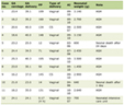 Click to view | Table 1. Pre-Treatment Clinical Stage and Post-Treatment Pathology Stage, Available for 35 Patients |
Examination of rectal biopsies and corresponding surgical resections
Two pathologists (DA and RM) assessed pre-treatment biopsies and the corresponding post-surgical specimens. In pre-treatment biopsies, they documented moderate to poorly differentiated adenocarcinomas in 66.6% of cases and moderately differentiated adenocarcinomas in 33.3% of cases. Post-treatment pathology (ypTNM) was staged for all 45 patients, revealing that the most frequent stage was ypT3N0 (33.3%), followed by ypT2N0 (17.8%). ypN0 nodal status was observed in 60% of patients, while 26.7% had ypN1, and 13.3% had ypN2 involvement. Metastasis (ypM1) was present in three patients (6.7%).
In 93.3% of patients, surgical margins were clear; anterior circumferential involvement was observed in 4.4% of cases, and posterior involvement was noted in 2.2%. Deposits were present in 11.1% of patients. TRS showed that three patients were TRS 1 (6.6%), 37 patients scored TRS 2 (82.2%), and five scored TRS 3 (11.1%). Extramural venous invasion was seen in four cases (8.8%) and LVI invasion in eight (17.7%).
Ki-67 and p53 expression in rectal adenocarcinoma in initial biopsy specimens and corresponding surgical resection
Strong Ki-67 staining was the predominant intensity in both the pre-treatment biopsies and the corresponding post-treatment specimens, although its frequency decreased from 37 (82.2%) to 32 (71.1%) cases following nCRT. The proportion of tumors showing Ki-67 expression in > 70% of tumor cells also decreased from 34 (75.5%) before treatment to 20 (44.4%) after treatment, whereas complete absence of staining increased from one (2.2%) to six (13.3%) cases (Table 2). A representative case demonstrating strong Ki-67 expression before nCRT and complete absence of expression in the corresponding post-treatment specimen is shown in Figure 1. Similarly, strong p53 staining remained the predominant intensity following treatment, although it decreased from 32 (71.1%) to 27 (60.0%) cases. The proportion of tumors showing p53 expression in > 70% of tumor cells decreased from 32 (71.1%) to 25 (55.5%), while complete absence of p53 staining increased from five (11.1%) to nine (20.0%) cases. The distribution of p53 IHC patterns shifted from predominantly wild-type before treatment, observed in 25 (55.6%) cases, to predominantly mutant-pattern after treatment, observed in 28 (62.2%) cases (Table 3).
Click to view | Table 2. Ki-67 Expression Before and After nCRT (N = 45) |
 Click for large image | Figure 1. Rectal adenocarcinoma in the same patient before and after neoadjuvant chemoradiotherapy (nCRT). (a) Strong Ki-67 expression in 90% of tumor cells before nCRT. (b) Post-nCRT surgical resection demonstrating complete absence of Ki-67 expression. |
Click to view | Table 3. p53 Expression Before and After nCRT (N = 45) |
Comparison of Ki-67 and p53 expression in rectal adenocarcinoma in initial biopsy specimens and corresponding surgical resection
The response to therapy showed that 82.2% of patients had a partial response (TRS 2), 11.1% had poor/no response (TRS 3), and 6.7% achieved a near complete response (TRS 1). Metastases were absent in 84.4% of patients, with post-treatment hepatic metastases seen in 6.7% of the sample.
The analysis of clinical and demographic factors in relation to therapy response revealed no significant associations. Age group did not appear to affect response to therapy (P = 0.741), and there was no significant difference in therapy response based on gender (P = 0.456). Also, the presence of a family history of cancer showed no clear impact on therapy response (P = 0.798).
For Ki-67, the mean percentage of staining also declined from 72.4% (pre-treatment) to 50.7% (post-treatment), with a mean difference of 32.9%. Strong staining remained predominant, though it decreased from 82.2% to 71.1%, while negative staining increased from 2.2% to 13.3% (Table 4).
Click to view | Table 4. Comparison of Ki-67 and p53 Expression in Rectal Adenocarcinoma in Initial Biopsy Specimens and Corresponding Surgical Resection |
When assessing the association between changes in p53 and Ki-67 expression and tumor response to therapy, the results varied by cutoff thresholds. For p53, using a 30% difference cutoff, the chi-square test showed no significant association with response to therapy (P = 0.419). At the 50% cutoff, the association approached significance (P = 0.088), but at the 70% cutoff, it remained nonsignificant (P = 0.437) (Table 5).
Click to view | Table 5. Association Between Changes in Ki-67 and p53 Expression and Tumor Response According to Different Cutoff Thresholds |
In contrast, Ki-67 showed a clearer association with tumor response. At the 30% cutoff, the association was not significant (P = 0.145), but it became statistically significant at the 50% cutoff (P = 0.019) and was highly significant at the 70% cutoff (P < 0.001) (Table 5). These findings indicate that larger reductions in Ki-67 expression were associated with pathological tumor response within this cohort, whereas comparable associations were not observed for p53 expression. Given the limited sample size and the marked imbalance between TRS groups, these associations should be interpreted cautiously.
However, analyzing the relationship between changes in p53 IHC pattern and tumor response, no significant association was found (P = 0.193). This indicates that shifts between mutant-pattern and wild-type-pattern p53 staining were not clearly linked to treatment outcomes.
In assessing whether reductions in Ki-67 or p53 were associated with therapy response, linear regression demonstrated significant differences in Ki-67 changes across response categories. Compared with TRS 1 (near-complete response), the estimated differences were −0.472 (95% CI, −0.761 to −0.183; P = 0.002) for TRS 2 and −0.447 (95% CI, −0.798 to −0.096; P = 0.014) for TRS 3. In contrast, no significant differences in p53 changes were observed for TRS 2 (estimate: −0.218; 95% CI, −0.577 to 0.141; P = 0.227) or TRS 3 (estimate: −0.347; 95% CI, −0.783 to 0.089; P = 0.117), compared with TRS 1. By examining the association of pre-treatment Ki-67 and p53 expression with therapy response, no significant associations were found. For Ki-67, the one-way ANOVA (P = 0.289) and the Kruskal–Wallis test (P = 0.509) both showed no significant association with pathological tumor response. Moreover, pre-treatment p53 staining showed no significant relationship with therapy response, with nonsignificant results in both the ANOVA (P = 0.741) and the Kruskal–Wallis test (P = 0.409).
Evaluating the association of Ki-67 and p53 with clinical and pathological features, mixed results were observed. Ki-67 expression was significantly linked to LVI (P < 0.001). However, no significant associations were found with tumor deposits (P = 0.172), extramural venous invasion (P = 0.459), or margin involvement (P = 0.984). For p53, none of these features showed a significant relationship, though margin involvement approached significance (P = 0.069).
Finally, regarding the Ki-67 pre-treatment staining intensity, no significant association was found with response to therapy or LVI.
For survival outcomes, at the 6-month follow-up, the OS rate was 95.6%, with two deaths recorded among the 45 patients. Similarly, the DFS rate was 95.6%, with two patients developing disease recurrence during the follow-up period.
| Discussion | ▴Top |
This study evaluated changes in Ki-67 and p53 expression in rectal adenocarcinoma following nCRT and explored their association with pathological tumor response. While pre-treatment expression levels of both markers were not associated with therapy response, Ki-67 expression decreased significantly following treatment (P < 0.001), with greater treatment-associated reductions associated with better pathological tumor response. These findings suggest that, in our cohort, the change in proliferative activity following nCRT may provide more information regarding treatment response than pre-treatment Ki-67 expression alone. The analysis of clinical and demographic factors in relation to therapy response revealed no significant associations. Age group did not appear to affect response to therapy (P = 0.741), and there was no significant difference in therapy response based on gender (P = 0.456). Similarly, the presence of a family history of colon cancer showed no clear impact on therapy response (P = 0.798).
Ki-67 and p53 have been investigated as potential biomarkers of response to neoadjuvant treatment in rectal cancer; however, previous findings have been inconsistent. Terzi et al found that pre-treatment expressions of Ki-67 and p53 did not significantly predict histopathological response to preoperative chemoradiotherapy [17]. In contrast, Jakob et al [18] evaluated Ki-67 and p53 expressions in pre-treatment biopsies and post-treatment resection specimens from patients with locally advanced rectal cancer receiving nCRT. Responders demonstrated significantly lower Ki-67 expression than non-responders in both pre- and post-treatment specimens, and Ki-67 expression correlated positively with thymidylate synthase (TS) mRNA levels. p53 expression was not associated with either tumor regression or TS expression, supporting Ki-67, but not p53, as a potential predictor of response to neoadjuvant therapy. In their cohort, pre-treatment and post-treatment specimens were available for 22 and 40 patients, respectively [18].
In another study, Rau et al [19] prospectively evaluated paired pre- and post-treatment tumor samples from 66 patients with rectal cancer to investigate dynamic changes in Ki-67, p53, p21, BAX, and hMSH2 following nCRT. An increase in Ki-67 expression after treatment and a decrease in p21 expression were associated with improved DFS, whereas pre-treatment p53 expression and p53 mutation status had no prognostic value. The authors concluded that dynamic treatment-induced biomarker changes may provide greater prognostic information than baseline expression, particularly highlighting p21 induction as a potential mechanism of treatment resistance.
Our findings therefore add to the existing literature by demonstrating that, although baseline Ki-67 expression was not associated with pathological response in our cohort, the magnitude of its treatment-associated reduction was associated with better pathological tumor response, supporting further investigation of dynamic changes in Ki-67 as a marker of treatment effect. In contrast, p53 expression showed no significant association with therapy response at either the pre- or post-treatment stage (ANOVA P = 0.741; Kruskal–Wallis P = 0.409). Although a modest reduction in p53 expression post-treatment was observed (P = 0.019), this did not translate into a significant correlation with clinical outcomes. These findings are consistent with previous studies, such as Terzi et al [17], which also found no predictive value of p53 expression in rectal cancer patients undergoing nCRT. Interestingly, p53 IHC patterns changed in some patients following treatment; however, these changes were not significantly associated with therapy response (P = 0.193). This suggests that p53 IHC patterns may not be a reliable indicator of treatment efficacy in this context.
When evaluating expression changes using cutoff thresholds, Ki-67 again demonstrated stronger clinical relevance, with a significant reduction in Ki-67 expression after treatment and a strong correlation in therapy response at higher cutoff values (50% and 70%). In contrast, p53 showed no significant associations at any threshold. Importantly, these cutoffs represent the magnitude of treatment-associated change in Ki-67 expression rather than absolute Ki-67 expression levels. The cutoff analyses were exploratory and were not intended to establish optimal or clinically validated thresholds.
Ki-67 has been considered a cellular proliferation marker, and its high expression has been associated with a poorer prognosis in rectal cancer. Yoshikawa et al found that patients with a high Ki-67 index had a 5-year survival rate of 68%, compared to 100% for those with a low Ki-67 index. This establishes the potential of Ki-67 as an independent prognostic marker in rectal cancer [20].
Lei et al identified a Ki-67 ≥ 60% cutoff as an independent biomarker for predicting both mortality and metastasis in CRC [1]. Similarly, Liu et al reported that Ki-67 expression above 50% was associated with improved DFS and therapeutic outcomes in patients receiving surgery and adjuvant chemotherapy, suggesting that high proliferative activity can increase tumor response to chemotherapy [9]. In contrast, Tong et al evaluated the prognostic significance of Ki-67 expression in 1,090 patients with CRC, stratifying absolute Ki-67 expression in 25% intervals within the AJCC-8 staging system [2]. Higher Ki-67 expression was associated with several adverse clinicopathological features, including greater invasive depth, poorer differentiation, advanced stage, and lymph node involvement, and multivariable analysis identified Ki-67 as an independent prognostic factor. The authors proposed 25% absolute Ki-67 expression as a useful classification cutoff, with higher Ki-67 expression associated with poorer prognosis [2]. However, the thresholds reported in these studies were based on absolute Ki-67 expression and should not be directly compared to the change-based cutoffs evaluated in the current study. Our research utilized cutoffs of 30%, 50%, and 70%, which are not absolute expressions but rather reflect the degree of changes from pre-treatment to post-treatment. Therefore, the clinical significance of these cutoffs cannot be inferred from the thresholds previously reported. Given the limited sample size and imbalance across tumor regression groups, the associations identified at the 50% and 70% cutoffs require validation in larger independent cohorts before any clinical application.
Associations of other pathological features, such as LVI, were found to be significant (P < 0.001) with Ki-67 in our study; direct studies specifically linking Ki-67 expression to LVI in CRC are limited. The high proliferative index indicated by Ki-67 could potentially correlate with the aggressive nature of tumors that exhibit LVI. The presence of LVI often indicates a more aggressive tumor phenotype, which could be reflected in higher Ki-67 expression levels, although this relationship is not explicitly detailed in the studies provided. The interplay between Ki-67 and LVI may thus serve as a valuable composite marker for stratifying patients by risk and guiding treatment decisions. Therefore, both high Ki-67 expression and LVI are independently associated with poor prognosis, indicating that they may contribute to tumor aggressiveness through different but potentially overlapping pathways [2, 21]. p53, however, showed no significant associations with LVI, tumor deposits, or margin involvement, although margin involvement approached significance (P = 0.069).
The reduction in Ki-67 expression following nCRT may reflect treatment-induced DNA damage, leading to cell-cycle arrest and apoptosis, while surviving resistant tumor cells may enter a quiescent or senescent state. p53 also plays an important role in the response to DNA damage through cell-cycle arrest, DNA repair, and apoptosis. However, mutations in p53 can hinder these protective mechanisms, allowing tumor cells to survive treatment and potentially leading to resistance. Although p53 expression was not significantly associated with pathological response in our study, its biological role in treatment response is well established. However, p53 IHC should be interpreted with caution, as it does not directly determine TP53 mutation status [22, 23]. In this study, the significant associations observed were limited to changes in Ki-67 expression following nCRT rather than baseline pre-treatment expression. Therefore, our findings support the role of changes in Ki-67 expression as a marker of treatment response rather than baseline Ki-67 expression as a validated pre-treatment predictive biomarker. Larger prospective studies incorporating pre-treatment biomarker assessment and long-term clinical outcomes are needed to determine the clinical utility of baseline Ki-67 expression and to validate the associations observed in our study.
Our study has several limitations, the most significant being the relatively small sample size of 45 participants with most patients classified as TRS 2, as many patients did not meet the inclusion criteria. However, it is important to highlight that the overall incidence of CRC in Jordan is relatively low compared with countries with higher incidence rates [24]. In addition, the evaluation of multiple Ki-67 cutoff thresholds was exploratory and should be interpreted cautiously until validated in larger, independent cohorts. The relatively short follow-up period (6 months) limited our ability to evaluate long-term oncologic outcomes, including local recurrence, DFS, and OS. Future studies with longer follow-up are warranted to determine whether Ki-67 and p53 expression are associated with long-term oncologic outcomes in addition to pathological tumor response. An additional limitation of this study is the exclusion of patients with a complete pathological response (TRS 0) from the paired biomarker analysis because no residual tumor was available for post-treatment immunohistochemical assessment. Furthermore, the pre-treatment data required to analyze baseline biomarker expressions in these patients were not available retrospectively. Future prospective studies that include all TRS categories are needed to validate these findings and further evaluate the clinical utility and optimal cutoff values for Ki-67 and p53.
Intratumoral heterogeneity may influence Ki-67 assessment, as pre-treatment biopsies may not fully represent the proliferative activity of the entire tumor. Post-treatment assessment may also be affected by treatment-related changes and the limited amount of residual viable tumor [25]. Moreover, potential selection bias related to the retrospective single-center design should also be considered. Moreover, molecular analyses, including TP53 sequencing and MSI/mismatch repair (MMR) assessment, were not performed and may have provided additional biological and prognostic information.
Despite these limitations, Ki-67 remains a practical proliferation marker in pathology. It is relatively cost-effective, widely accessible, and can be assessed using standard IHC techniques. Unlike molecular assays, Ki-67 assessment does not require advanced molecular laboratory facilities, making it potentially useful in resource-limited settings [26].
Although p53 IHC is widely used in diagnostic pathology as a surrogate marker for TP53 alterations, it is not a direct substitute for molecular TP53 sequencing, as discordance between IHC staining patterns and mutation status may occur. Therefore, p53 IHC should be interpreted cautiously as a surrogate marker of TP53 status [27].
Our findings warrant larger prospective studies to validate these associations and further explore the integration of Ki-67 into routine pathological assessment. While p53 remains biologically relevant, no significant association with pathological tumor response was observed in our cohort. Future research should also investigate molecular subtypes and emerging biomarkers to refine personalized treatment strategies for rectal adenocarcinoma.
Conclusions
Overall, our findings suggest that changes in Ki-67 expression following nCRT are associated with pathological tumor response, whereas pre-treatment Ki-67 and p53 expression were not. This supports the role of changes in Ki-67 expression as a marker associated with treatment response, which may also provide complementary information on the biological response of the residual tumor following nCRT, beyond the morphological changes assessed by the TRS. However, as patients with complete pathological response (TRS 0) were excluded because no viable tumor was available for post-treatment assessment, these findings are limited to patients with residual viable tumor and should not be generalized to the full spectrum of pathological response.
Although pre-treatment Ki-67 expression was not associated with pathological response in our cohort, its assessment remains of interest given the conflicting findings reported in the literature. Larger prospective studies incorporating pre-treatment biomarker assessment and long-term clinical outcomes are needed to further define the clinical utility of baseline Ki-67 expression and to validate the associations observed in our study.
Acknowledgments
None to declare.
Financial Disclosure
This work was supported by an intramural fund from KHCC, Jordan.
Conflict of Interest
The authors declare that they have no conflict of interest.
Informed Consent
The requirement for informed consent was waived by the IRB due to the retrospective nature of the study and the use of deidentified data.
Author Contributions
DA and RM conducted the clinical data search, reviewed the pathology sections, and reported the TRSs and diagnoses for each case, and drafted the initial manuscript. MA, MS and FB assisted in retrieving the pathology blocks, performing the immunostaining, and securing IRB approval and funding. BA and LA performed the statistical analysis and drafted the results section. NH revised the manuscript and did English editing. AA and SA wrote the discussion section of the manuscript. MS, MQ, and NH contributed to writing, editing, and finalizing the manuscript, and obtained the pathology images.
Data Availability
The datasets generated and analyzed during the current study are not publicly available due to institutional and patient confidentiality restrictions but are available from the corresponding author on reasonable request, subject to the IRB approval.
AI Use Declaration
No AI tools were used in the design, data collection, data analysis, interpretation of results, or preparation of this manuscript. All analyses, interpretations, and writing were performed by the authors.
| References | ▴Top |
- Lei HT, Yan S, He YH, Xu N, Zhao M, Yu CJ, Li HL, et al. Ki67 testing in the clinical management of patients with non-metastatic colorectal cancer: Detecting the optimal cut-off value based on the Restricted Cubic Spline model. Oncol Lett. 2022;24(6):420.
doi pubmed - Tong G, Zhang G, Liu J, Zheng Z, Chen Y, Niu P, Xu X. Cutoff of 25% for Ki67 expression is a good classification tool for prognosis in colorectal cancer in the AJCC-8 stratification. Oncol Rep. 2020;43(4):1187-1198.
doi pubmed - Barman D, Bhattacharjee P, Das A, et al. Ki-67 expression in colorectal cancer: Correlation with clinicopathological features. J Gastrointest Oncol. 2021;12(3):1234-1242.
- Ashar A, Patel M. Ki-67 as a prognostic marker in colorectal cancer: A retrospective study. Indian J Pathol Microbiol. 2016;59(4):451-456.
- Bhagyalakshmi A, Sreelekha A, Kasi Babu A, et al. Ki-67 proliferation index and clinicopathological patterns in colorectal carcinomas. J Clin Sci Res. 2015;4(2):85-91.
doi - Yuan C, Huang J, Wang Y, Xiao H. Exploration and validation of the Ki67, Her-2, and mutant P53 protein-based risk model, nomogram and lymph node metastasis model for predicting colorectal cancer progression and prognosis. Front Oncol. 2023;13:1236441.
doi pubmed - Yassen NN, Abouelfadl DM, Abbas NF, Soliman ASA, Shabana ME. Microsatellite instability screening in colorectal carcinoma: immunohistochemical analysis of MMR proteins in correlation with clinicopathological features and Ki-67 protein expression. Bull Natl Res Cent. 2023;47:155.
doi - Luo ZW, Zhu MG, Zhang ZQ, Ye FJ, Huang WH, Luo XZ. Increased expression of Ki-67 is a poor prognostic marker for colorectal cancer patients: a meta analysis. BMC Cancer. 2019;19(1):123.
doi pubmed - Liu Q, Ran D, Wang L, Feng J, Deng W, Mei D, Peng Y, et al. Association between Ki67 expression and therapeutic outcome in colon cancer. Oncol Lett. 2023;25(6):272.
doi pubmed - Gao Z, Cao H, Xu X, Wang Q, Wu Y, Lu Q. Prognostic value of lymphovascular invasion in stage II colorectal cancer patients with an inadequate examination of lymph nodes. World J Surg Oncol. 2021;19(1):125.
doi pubmed - Huh JW, Kim HR, Kim YJ. Prognostic value of lymphovascular invasion in patients with stage I colorectal cancer. Int J Colorectal Dis. 2013;28(3):375-380.
doi - Sauer R, Liersch T, Merkel S, Fietkau R, Hohenberger W, Hess C, Becker H, et al. Preoperative versus postoperative chemoradiotherapy for locally advanced rectal cancer: results of the German CAO/ARO/AIO-94 randomized phase III trial after a median follow-up of 11 years. J Clin Oncol. 2012;30(16):1926-1933.
doi pubmed - Lynch S, Mirza T, Lateef F, Hassan S. The importance of immunohistochemical evaluation of Ki-67 in detecting early malignant changes in colorectal adenomatous polyps. Med Forum. 2024;35(1):28-32.
doi - Brodsky JT, Cohen AM. Lymphovascular invasion in colorectal cancer: A prognostic indicator. Cancer. 1992;70(11):2281-2286.
doi - Loughrey MB, Quirke P, Shepherd NA; Royal College of Pathologists Cancer Services Working Group. G049 Dataset for histopathological reporting of colorectal cancer v5. Royal College of Pathologists; 2023. Available from: https://www.rcpath.org/resourceLibrary/g049-dataset-for-histopathological-reporting-of-colorectal-cancer.html.
- Ryan R, Gibbons D, Hyland JM, Treanor D, White A, Mulcahy HE, O'Donoghue DP, et al. Pathological response following long-course neoadjuvant chemoradiotherapy for locally advanced rectal cancer. Histopathology. 2005;47(2):141-146.
doi pubmed - Terzi C, Canda AE, Sagol O, Atila K, Sonmez D, Fuzun M, Gorken IB, et al. Survivin, p53, and Ki-67 as predictors of histopathologic response in locally advanced rectal cancer treated with preoperative chemoradiotherapy. Int J Colorectal Dis. 2008;23(1):37-45.
doi pubmed - Jakob C, Liersch T, Meyer W, Becker H, Baretton GB, Aust DE. Predictive value of Ki67 and p53 in locally advanced rectal cancer: correlation with thymidylate synthase and histopathological tumor regression after neoadjuvant 5-FU-based chemoradiotherapy. World J Gastroenterol. 2008;14(7):1060-1066.
doi pubmed - Rau B, Sturm I, Lage H, Berger S, Schneider U, Hauptmann S, Wust P, et al. Dynamic expression profile of p21WAF1/CIP1 and Ki-67 predicts survival in rectal carcinoma treated with preoperative radiochemotherapy. J Clin Oncol. 2003;21(18):3391-3401.
doi pubmed - Yoshikawa K, Shimada M, Higashijima J, Nakao T, Nishi M, Takasu C, Kashihara H, et al. Ki-67 and survivin as predictive factors for rectal cancer treated with preoperative chemoradiotherapy. Anticancer Res. 2018;38(3):1735-1739.
doi pubmed - Yahyazadeh H, Mafi A R, Khatooni E, Beheshti M, Abdollahinejad A. Lymphovascular and perineural invasions are independently associated with advanced colorectal carcinoma. Int J Cancer Manag. 2019;12(11):e95540.
doi - Huang P, Zhao X, Fei H, Zhang L, Liu X, Yu Y, Wu R, et al. Watch-and-Wait strategy for locally advanced rectal cancer after neoadjuvant chemoradiotherapy: a comprehensive review. Front Oncol. 2026;16:1760042.
doi pubmed - Li W, Li L, Yang H, Shi C, Lei Z, Guo L, Wang Y. Unraveling the role of TP53 in colorectal cancer therapy: from wild-type regulation to mutant. Front Biosci (Landmark Ed). 2024;29(7):272.
doi pubmed - Eser S, Chang J, Charalambous H, Silverman B, Demetriou A, Yakut C, Nimri O, et al. Incidence patterns of colorectal cancers in four countries of the Middle East Cancer Consortium (Cyprus, Jordan, Israel, and Izmir, Turkey) compared with those in the United States Surveillance, Epidemiology, and End Results Program. Turk J Gastroenterol. 2018;29(1):36-44.
doi pubmed - Kressner U, Lindmark G, Gerdin B, Pahlman L, Glimelius B. Heterogeneity in proliferation markers in colorectal cancer. Anticancer Res. 1995;15(6B):2755-2761.
pubmed - Kasprzak A. Prognostic biomarkers of cell proliferation in colorectal cancer (CRC): from immunohistochemistry to molecular biology techniques. Cancers (Basel). 2023;15(18):4570.
doi pubmed - Osakabe M, Yamada N, Sugimoto R, Uesugi N, Nakao E, Honda M, Yanagawa N, et al. The pattern-based interpretation of p53 immunohistochemical expression as a surrogate marker for TP53 mutations in colorectal cancer. Virchows Arch. 2025;486(2):333-341.
doi pubmed
This article is distributed under the terms of the Creative Commons Attribution 4.0 International License (CC BY 4.0), which permits unrestricted use, distribution, and reproduction in any medium, including commercial use, provided the original work is properly cited.
World Journal of Oncology is published by Elmer Press Inc.