| World Journal of Oncology, ISSN 1920-4531 print, 1920-454X online, Open Access |
| Article copyright, the authors; Journal compilation copyright, World J Oncol and Elmer Press Inc |
| Journal website https://wjon.elmerpub.com |
Case Report
Volume 17, Number 6, December 2026, pages 923-939

Metastatic Triple-Negative Male Breast Carcinoma of Occult Primary With Axillary Nodal and Cutaneous Metastases
Emiri Sugiyamaa, b, Elise Di Lenac, Kana Matsudaa, Saki Andoa, Junko Takaia, Seiji Sugiyamad, Fumio Sakashitaa, b, Kazuya Yamaguchib, Makoto Takeuchia, Kazuaki Takabec, Yoshihisa Tokumarua, e
aDepartment of Breast Surgery, Central Japan International Medical Center, Minokamo, Gifu, Japan
bDepartment of Gastroenterological Surgery, Central Japan International Medical Center, Minokamo, Gifu, Japan
cDepartment of Surgical Oncology, Roswell Park Comprehensive Cancer Center, Buffalo, NY, USA
dDepartment of Pathology, Central Japan International Medical Center, Minokamo, Gifu, Japan
eCorresponding Author: Yoshihisa Tokumaru, Department of Breast Surgery, Central Japan International Medical Center, Minokamo, Gifu, Japan
Manuscript submitted July 7, 2026, accepted September 16, 2026, published online November 11, 2026
Short title: Metastatic Breast Carcinoma of Occult Primary
doi: https://doi.org/10.14740/wjon2844
| Abstract | ▴Top |
Classical occult breast cancer (OBC), defined as T0N+M0 breast cancer presenting with axillary lymph node metastasis without an identifiable primary breast lesion, is exceptionally rare in men. A 77-year-old man was admitted to the emergency department with hypothermia, and contrast-enhanced computed tomography performed during the initial evaluation revealed enlarged left axillary lymph nodes and cutaneous nodules on the left chest wall. No obvious primary lesion was identified. Because malignant lymphoma or metastasis from another primary malignancy was initially suspected, biopsy of a cutaneous nodule was performed. Histopathological examination demonstrated infiltration of atypical carcinoma cells from the dermis to the subcutaneous tissue. Immunohistochemical analysis was consistent with a mammary origin. Estrogen and progesterone receptors were negative, and human epidermal growth factor receptor 2 expression was negative by fluorescence in situ hybridization, consistent with a triple-negative subtype. Further diagnostic workup identified no primary breast or other malignancy. Axillary lymph node dissection was performed for diagnostic confirmation and regional disease control. The dissected lymph nodes showed tumor morphology and immunophenotype similar to those of the cutaneous lesion, further supporting a diagnosis of mammary origin. The multiple cutaneous lesions were clinically interpreted as metastases; accordingly, the disease was classified as cT0N1M1, stage IV. Germline BRCA1/2 testing revealed no pathogenic variants. The patient was diagnosed with metastatic triple-negative breast carcinoma of occult primary with axillary nodal and cutaneous involvement. Because programmed death-ligand 1 (PD-L1) expression was positive, systemic therapy with pembrolizumab, gemcitabine, and carboplatin was initiated. The clinical course after treatment initiation was initially stable; however, the patient developed recurrent severe hypothermia after the second cycle and died approximately 6 months after the initial presentation. The precise cause of the terminal event remained undetermined, and an immune-related endocrine adverse event could not be excluded because thyroid and pituitary-adrenal function were not evaluated at the terminal admission. This case highlights the importance of comprehensive immunohistochemical evaluation in diagnosing metastatic breast carcinoma of occult primary when no primary breast lesion is detected by imaging. It also highlights the feasibility of PD-L1 testing and combined positive score (CPS)-guided treatment selection in metastatic triple-negative breast carcinoma of occult primary.
Keywords: Occult male breast cancer; Triple-negative breast cancer; Axillary lymph node metastasis; Cutaneous metastasis
| Introduction | ▴Top |
Male breast cancer is rare, accounting for approximately 1% of all breast cancers [1, 2]. Classical occult breast cancer (OBC), traditionally defined as T0N+M0 breast cancer presenting with axillary lymph node metastasis without an identifiable primary breast lesion, accounts for approximately 0.3–1.0% of all breast cancers [3]. When this presentation occurs in men, it is referred to as occult male breast cancer (OMBC), an exceedingly rare entity with only sporadic cases reported worldwide [4, 5]. However, rare male cases presenting with non-axillary extramammary metastases without an identifiable breast primary have also been reported. Therefore, for the purposes of the present literature review, we broadened the eligibility criteria to include reported cases of male breast carcinoma of occult primary presenting with axillary nodal and/or other extramammary metastatic disease without an identifiable breast primary on initial clinical and imaging evaluation.
OMBC poses significant diagnostic challenges because patients typically present with isolated axillary lymphadenopathy, leading initially to suspicion of lymphoma or metastatic disease from another primary site. Conventional imaging modalities, including mammography, ultrasonography, and magnetic resonance imaging (MRI), frequently fail to identify a primary breast lesion [6]. Therefore, pathological evaluation with immunohistochemical analysis plays a pivotal role in establishing the diagnosis. Immunostaining for markers such as cytokeratin (CK)7, GATA binding protein 3 (GATA3), mammaglobin, gross cystic disease fluid protein 15 (GCDFP-15), estrogen receptor (ER), progesterone receptor (PR), and human epidermal growth factor receptor 2 (HER2) is useful in supporting a mammary origin [7, 8]. However, these markers should be interpreted as part of a broader diagnostic panel, because GATA3 and other breast-associated markers may also be expressed in sweat gland and cutaneous adnexal tumors. Therefore, additional markers, including p63 and CK5/6, as well as clinicoradiological findings, are important for excluding non-mammary primary tumors [9, 10].
Due to the extreme rarity of OMBC, no disease-specific diagnostic or therapeutic guidelines have been established. Current clinical practice is therefore largely extrapolated from treatment strategies for female breast cancer or from the National Comprehensive Cancer Network (NCCN) guidelines for cancer of unknown primary (CUP), which recommend tailoring therapy based on the most likely site of origin inferred from histopathology and immunohistochemical [11]. Reported cases indicate that hormone receptor–positive OMBC has been treated with endocrine therapy [12, 13], whereas HER2-positive OMBC may benefit from HER2-targeted treatment [14, 15].
Given the paucity of reported cases, each additional report provides valuable insight into the clinical characteristics and optimal management of this rare disease. Herein, we report a rare case of metastatic triple-negative breast carcinoma of occult primary in a male patient presenting with axillary lymphadenopathy and cutaneous metastases, and we review the published literature with a focus on diagnostic challenges, immunohistochemical evaluation, and therapeutic strategies.
| Case Report | ▴Top |
A 77-year-old man was admitted to the emergency department with hypothermia. Contrast-enhanced computed tomography (CT) performed during the initial evaluation revealed enlarged left axillary lymph nodes and cutaneous nodules on the left lower lateral anterior chest wall (Fig. 1). No obvious primary lesion was identified. Because malignant lymphoma was initially suspected, a biopsy of the cutaneous nodule was performed.
 Click for large image | Figure 1. (a) Contrast-enhanced computed tomography findings showing enlarged left axillary lymph nodes (arrow). (b) Clinical photograph of multiple erythematous papular lesions on the left anterior chest wall suspicious for cutaneous metastases (arrow). |
Histopathological examination demonstrated poorly differentiated carcinoma cells with prominent nucleoli arranged in trabecular and nested patterns. Immunohistochemical analysis showed positivity for AE1/AE3, CAM5.2, CK7, mammaglobin, GATA3, and GCDFP-15, and negativity for CK20, p63, carcinoembryonic antigen (CEA), caudal-type homeobox 2 (CDX2), napsin A/thyroid transcription factor 1 (TTF-1), chromogranin A, CD45, synaptophysin, human melanoma black 45 (HMB45), and melanoma antigen recognized by T cells 1 (Melan-A) (Fig. 2). These findings suggested cutaneous metastasis of breast carcinoma, although sweat gland or sweat duct carcinoma could not be completely excluded. The immunohistochemical profile of the index case and its diagnostic interpretation are summarized in Table 1. The patient was therefore referred to our department for further examination. Because the differential diagnosis included a primary cutaneous adnexal (sweat gland) carcinoma, the patient was also evaluated by dermatologists. Based on the absence of a clinically apparent primary adnexal lesion, a primary sweat gland carcinoma was considered clinically unlikely. Immunohistochemical analysis demonstrated negative ER (clone SP1) and PR (clone 1E2) expression, with < 1% nuclear staining for both markers, and equivocal HER2 expression (2+). HER2 fluorescence in situ hybridization (FISH) showed no amplification, with a HER2/CEP17 ratio of 0.8 and an average HER2 copy number of 1.4 signals per cell. The Ki-67 labeling index was 28%. Based on the negative ER and PR expression and absence of HER2 amplification, the tumor was classified as triple-negative. Additional immunohistochemical analysis of the available tumor tissue demonstrated androgen receptor (AR) positivity and SRY-box transcription factor 10 (SOX10) negativity. CK5/6 was focally positive in a small subset of tumor cells. Immunohistochemical staining for trichorhinophalangeal syndrome type 1 (TRPS1) could not be performed because the antibody was not available at our institution.
 Click for large image | Figure 2. Histopathological and immunohistochemical findings of the cutaneous metastatic lesion. (a) Hematoxylin and eosin staining showing poorly differentiated carcinoma cells with prominent nucleoli arranged in trabecular and nested patterns in the dermis and subcutaneous tissue (original magnification, × 40; scale bar = 500 µm). (b) Estrogen receptor (ER; clone SP1) showing negative expression (original magnification, × 40; scale bar = 500 µm). (c) Progesterone receptor (PR; clone 1E2) showing negative expression (original magnification, × 40; scale bar = 500 µm). (d) Human epidermal growth factor receptor 2 (HER2; clone 4B5) showing equivocal expression (2+) (original magnification, × 40; scale bar = 500 µm). (e) Cytokeratin 7 (CK7; clone SP52) showing positive expression (original magnification, × 40; scale bar = 500 µm). (f) Cytokeratin 20 (CK20; clone SP33) showing negative expression (original magnification, × 40; scale bar = 500 µm). (g) GATA binding protein 3 (GATA3; clone L50-823) showing positive expression (original magnification, × 40; scale bar = 500 µm). (h) Gross cystic disease fluid protein 15 (GCDFP-15; clone EP1582Y) showing positive expression (original magnification, × 40; scale bar = 500 µm). |
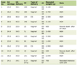 Click to view | Table 1. Immunohistochemical Profile of the Index Case and Its Diagnostic Interpretation |
Mammography and breast ultrasonography demonstrated no intramammary lesions. However, a lobulated hypoechoic mass measuring approximately 28 mm was identified in the left axilla, suggestive of lymph node metastasis. Breast MRI showed no abnormal enhancement within either breast. Serum prostate-specific antigen (PSA) was within the reference range. Positron emission tomography–computed tomography (PET-CT) demonstrated fluorodeoxyglucose (FDG) uptake in the left axillary lymph nodes, cutaneous and subcutaneous lesions of the left chest wall, parotid gland, and intestinal tract, whereas no abnormal FDG uptake was observed in the prostate. Subsequent otolaryngological evaluation, including MRI of the parotid gland, revealed no evidence of a primary salivary gland malignancy. Upper gastrointestinal endoscopy, colonoscopy, and small-bowel endoscopy also demonstrated no evidence of gastrointestinal malignancy (Fig. 3)
 Click for large image | Figure 3. Positron emission tomography–computed tomography (PET-CT) findings. (a) Increased fluorodeoxyglucose (FDG) uptake in the left axillary lymph nodes (arrow). (b) Increased FDG uptake in cutaneous and subcutaneous lesions of the left chest wall (arrow). (c) Increased FDG uptake in the parotid gland (arrow). (d) Increased FDG uptake in the pelvic intestinal tract (arrow). |
Because the primary site remained uncertain despite extensive imaging studies, axillary lymph node dissection (ALND) was subsequently performed for pathological confirmation and regional disease control. Histopathological examination of the dissected axillary lymph nodes revealed metastatic carcinoma in all seven excised lymph nodes (7/7), with morphological features similar to those observed in the initial skin biopsy. No mammary gland tissue or identifiable primary lesion was detected in the surgical specimen. Immunohistochemical analysis demonstrated positivity for CK7, GATA3, GCDFP-15, and mammaglobin, and negativity for CK20, further supporting mammary differentiation, although a primary cutaneous adnexal carcinoma could not be completely excluded (Fig. 4). Mastectomy was not performed because no primary breast lesion was identified on imaging studies and the primary site remained uncertain at the time of surgery.
 Click for large image | Figure 4. Histopathological and immunohistochemical findings of the axillary lymph node metastasis. (a) Hematoxylin and eosin staining showing metastatic carcinoma involving the axillary lymph node, with solid nests and sheet-like growth of atypical carcinoma cells (original magnification, × 40; scale bar = 500 µm). (b) Estrogen receptor (ER; clone SP1) showing negative expression (original magnification, × 40; scale bar = 500 µm). (c) Progesterone receptor (PR; clone 1E2) showing negative expression (original magnification, × 40; scale bar = 500 µm). (d) Human epidermal growth factor receptor 2 (HER2; clone 4B5) showing equivocal expression (2+) (original magnification, × 40; scale bar = 500 µm). (e) Cytokeratin 7 (CK7; clone SP52) showing positive expression (original magnification, × 40; scale bar = 500 µm). (f) Cytokeratin 20 (CK20; clone SP33) showing negative expression (original magnification, × 40; scale bar = 500 µm). (g) GATA binding protein 3 (GATA3; clone L50-823) showing positive expression (original magnification, × 40; scale bar = 500 µm). (h) Gross cystic disease fluid protein 15 (GCDFP-15; clone EP1582Y) showing positive expression (original magnification, × 40; scale bar = 500 µm). |
Following ALND, new cutaneous lesions developed on the left chest wall and subsequently progressed, prompting additional imaging to reassess disease extent and evaluate for distant metastases. PET-CT revealed FDG uptake in the left subclavian lymph nodes and chest wall, indicating disease progression. Because the chest wall skin lesions presented as separate dermal/subcutaneous nodules rather than direct extension from an identifiable breast primary, they were interpreted as cutaneous metastases after multidisciplinary discussion. No identifiable primary lesion was detected in the breast, and the chest-wall cutaneous lesions were clinically interpreted as cutaneous metastases. On this basis, the disease was classified as cT0N1M1, corresponding to stage IV breast cancer. Histopathological examination after ALND revealed metastatic carcinoma in all seven excised axillary lymph nodes (7/7), corresponding to pN2a. Because no breast resection was performed and no primary breast lesion was pathologically identified, a complete pathological TNM classification and pathological stage group could not be assigned.
Based on the clinical, radiological, and pathological findings, the patient was diagnosed with metastatic triple-negative breast carcinoma of occult primary with axillary nodal and cutaneous involvement. Given the diagnosis of male breast cancer, germline genetic testing was performed and was limited to BRCA1/2, with no pathogenic variants identified. Programmed death-ligand 1 (PD-L1) immunohistochemistry was performed using the 22C3 antibody clone and evaluated using the combined positive score (CPS); the tumor showed a CPS of ≥ 10. The specific staining platform could not be confirmed from the available records. Microsatellite instability (MSI) was assessed using the MSI Test Kit (FALCO), a multiplex polymerase chain reaction (PCR)–fragment analysis assay, and MSI-high was not detected. Systemic therapy with pembrolizumab in combination with gemcitabine and carboplatin was initiated for metastatic triple-negative breast cancer (TNBC). Because of the progression of the cutaneous metastatic disease, neither radiotherapy nor additional surgery was planned at that time, and systemic therapy was prioritized. Local therapy, including radiotherapy, could have been reconsidered depending on the subsequent response to systemic therapy and the extent of residual disease. The initial clinical course was stable without notable adverse events. However, after the second cycle of therapy, the patient presented to the emergency department with severe hypothermia and impaired consciousness. On the day of admission, he had been in his usual state of health until the afternoon, when he developed chills followed by impaired consciousness. His body temperature was 27 °C on arrival at the emergency department, and marked bradycardia was noted during transport. Despite active rewarming, progressive bradycardia and agonal respiration developed, followed by cardiopulmonary arrest. In accordance with the wishes of the patient’s family, cardiopulmonary resuscitation was not performed, and the patient died shortly thereafter.
The precise cause of the recurrent severe hypothermia remained undetermined. Notably, the patient had experienced an episode of severe hypothermia before pembrolizumab therapy. At the initial admission, thyroid-stimulating hormone (TSH), free triiodothyronine (T3), free thyroxine (T4), and adrenocorticotropic hormone (ACTH) were within the reference ranges, whereas serum cortisol was mildly elevated rather than decreased. Serum sodium, glucose, and creatine kinase (CK) were also within the reference ranges. Thyroid autoantibodies were not measured. These findings did not support overt hypothyroidism or adrenal insufficiency as the cause of the initial episode; however, no alternative cause was identified, and its etiology remained undetermined. The hypothermia subsequently resolved, and the patient’s body temperature had returned to the normal range by the time pembrolizumab-based therapy was initiated.
Thyroid and adrenal function were reassessed before pembrolizumab initiation and monitored during treatment, with TSH, free T3, free T4, ACTH, and cortisol remaining within the reference ranges. Repeat testing approximately 2 weeks before the terminal admission also showed no evidence of thyroid or adrenal dysfunction. However, TSH, free T4, cortisol, and ACTH were not measured at the terminal admission. Empirical hydrocortisone or levothyroxine was not administered. Therefore, myxedema coma, hypophysitis, or adrenal insufficiency related to immune checkpoint inhibitor therapy could not be definitively excluded. An autopsy was not performed.
This case report was prepared in accordance with the Case Report (CARE) reporting guideline. The clinical course is summarized in the following timeline.
Timeline of the case
June 2023 – The patient was admitted with severe hypothermia. CT revealed left axillary lymphadenopathy and multiple cutaneous and subcutaneous nodules on the left chest wall, without an identifiable primary lesion. The cause of the hypothermia remained undetermined.
June 2023 – Biopsy of a cutaneous nodule on the left chest wall demonstrated poorly differentiated carcinoma. The morphological and immunophenotypic findings supported mammary differentiation, although a primary cutaneous adnexal carcinoma could not be completely excluded.
July 2023 – PET-CT, mammography, breast ultrasonography, breast MRI, endoscopic examinations, and other investigations did not identify a definite primary tumor.
October 2023 – Left axillary lymph node dissection and excision of the chest-wall skin lesions were performed. Pathological findings supported triple-negative breast carcinoma of occult primary with axillary nodal and cutaneous involvement.
November 2023 – Postoperative PET-CT demonstrated disease progression in the left subclavian lymph nodes and chest wall. Pembrolizumab-based systemic therapy was initiated based on PD-L1 expression with a CPS of ≥ 10.
December 2023 – The second cycle of pembrolizumab-based therapy was administered.
December 2023 – Approximately 2 weeks after initiation of the second cycle, the patient presented to the emergency department with recurrent severe hypothermia, bradycardia, and impaired consciousness and died shortly thereafter. The cause of the terminal event remained undetermined, and an immune-related endocrine adverse event could not be definitively excluded.
| Literature Review | ▴Top |
Literature search and eligibility criteria
A structured literature search was performed using PubMed and Scopus from database inception through August 13, 2026. The reference lists of eligible articles and relevant reviews were also hand-searched to identify additional reports. The search strategy combined terms related to male sex, breast cancer, and occult or unknown primary disease. The complete search strategies for each database are provided (Supplementary Material 1, wjon.elmerpub.com).
For the purpose of this review, eligible cases included male breast carcinoma of occult primary presenting with axillary nodal and/or other extramammary metastatic disease without an identifiable breast primary on initial clinical and imaging evaluation. Case reports and case series providing individual patient-level data were eligible for inclusion. Reports involving female patients, accessory breast carcinoma, an identifiable breast primary at the initial evaluation, non-breast primary malignancies, duplicate patient reports, or insufficient individual clinical data were excluded. No language restrictions were applied. Two reviewers independently screened the titles and abstracts and subsequently assessed potentially eligible full-text articles, with disagreements resolved by consensus.
The database searches identified 57 records (PubMed, n = 29; Scopus, n = 28). After removal of 20 duplicate records, 37 records underwent title and abstract screening, of which 14 were excluded. Twenty-three full-text reports were assessed for eligibility, and one duplicate patient report was excluded. Twenty-two reports were included from the database searches. Four additional reports were identified through hand-searching of reference lists, resulting in a total of 26 reports describing 27 patients. The study-selection process is summarized in a Preferred Reporting Items for Systematic Reviews and Meta-Analyses (PRISMA)-style flow diagram (Supplementary Material 2, wjon.elmerpub.com).
Epidemiology and clinical characteristics
Male breast cancer is rare, accounting for approximately 1% of all breast cancers [1, 2]. OBC, described in early reports by Halsted and Cameron in the early 20th century, is defined as axillary lymph node metastasis from breast carcinoma in the absence of an identifiable primary breast lesion [16, 17]. When this condition occurs in men, it is referred to as OMBC, which is even rarer and represents a diagnostic and therapeutic challenge because of its atypical presentation [4, 5].
Rare male cases presenting with non-axillary extramammary metastases without an identifiable breast primary have also been reported. A previous systematic review by Gomez Garnica et al identified 11 case reports comprising 12 male patients with OBC, most of whom were diagnosed with advanced-stage disease, with substantial heterogeneity in receptor status and treatment strategies [18]. The present updated review identified 26 reports describing 27 patients, including more recently reported cases with non-axillary presentations and diverse molecular subtypes. The clinicopathological characteristics of the included cases are summarized in Table 2 [4, 12–15, 18–38].
Click to view | Table 2. Summary of Previously Reported Cases of Occult Male Breast Cancer Identified Through the Systematic Literature Review |
Among the 27 patients included in the present review, the median age was 65.5 years (range, 29–84 years) among the 26 patients for whom an exact age was reported. Based on the reported ER, PR, and HER2 results, six patients (22.2%) had hormone receptor-positive/HER2-negative disease, two (7.4%) had hormone receptor-positive disease with unreported HER2 status, three (11.1%) had hormone receptor-positive/HER2-positive disease, seven (25.9%) had hormone receptor-negative/HER2-positive disease, eight (29.6%) had TNBC, and the subtype remained unclear in one patient (3.7%). The triple-negative classification of the case reported by Su et al was based on the authors’ final interpretation despite discordant receptor findings between the axillary and pulmonary lesions [37].
After normalization of the stage notation, 11 patients (40.7%) were identified as having stage IV disease. Seven patients (25.9%) had explicitly documented non-stage IV disease, whereas stage could not be definitively categorized from the information provided in Table 2 [4, 12–15, 18–38] for the remaining nine patients (33.3%). The latter group includes the case reported by Akhtar et al [38], for which a post-neoadjuvant pathological stage of ypT0N1a was provided but the M category and overall stage group were not explicitly reported.
Among the 11 patients with stage IV disease, one had hormone receptor-positive/HER2-negative disease, one had hormone receptor-positive disease with unreported HER2 status, two had hormone receptor-positive/HER2-positive disease, four had hormone receptor-negative/HER2-positive disease, and three had TNBC.
Surgical approaches varied substantially. Mastectomy with or without axillary surgery was performed in nine patients (33.3%), whereas seven (25.9%) underwent axillary surgery without mastectomy. Four patients (14.8%) underwent diagnostic biopsy alone, six (22.2%) underwent no surgery, and one (3.7%) underwent resection of a metastatic lesion without breast or axillary surgery. Chemotherapy was administered in 24 patients (88.9%), radiotherapy in 12 (44.4%), endocrine therapy in nine (33.3%), HER2-targeted therapy in seven (25.9%), and immunotherapy in one (3.7%).
Clinical outcomes among the 11 patients with stage IV disease varied widely, ranging from prolonged disease control to death from progressive disease. Among the six patients with HER2-positive stage IV disease, including both hormone receptor-positive and hormone receptor-negative tumors, two were alive with disease, one achieved a complete response with 33 months of follow-up, one had stable disease at 48 months, one died of disease after 56 months, and the outcome was not reported in one. Among the three patients with stage IV triple-negative disease, one had no recurrence reported at 12 months, one was alive with disease, and one died of disease. The remaining two patients had hormone receptor-positive disease: one had no evidence of disease at 36 months, whereas the other transitioned to hospice care. Given the small number of cases, heterogeneity in metastatic burden and treatment, and variable follow-up duration, these findings should be interpreted descriptively, and no firm conclusions regarding prognostic differences among molecular subtypes can be drawn.
A numerical follow-up duration was reported for 15 patients. Using the numerical values provided in the reports, the median reported follow-up was 33 months, with values ranging from 4 to 56 months. These calculations included three qualified durations reported as approximately 48 months.
OBC is most commonly suspected when patients present with axillary lymphadenopathy and no detectable breast lesion on physical examination or imaging [39]. In this setting, imaging studies such as mammography, ultrasonography, MRI, and PET-CT are useful mainly for confirming the absence of an identifiable breast primary and excluding other primary malignancies, and assessing disease extent, rather than for establishing the diagnosis itself [6, 40]. A systematic review by de Bresser et al reported that breast MRI identified an otherwise occult breast lesion in approximately 72% of patients, highlighting its diagnostic value in this setting [40]. Therefore, histopathological and immunohistochemical evaluation remains essential for supporting a mammary origin.
Because disease-specific diagnostic and therapeutic guidelines are lacking, treatment strategies have generally been extrapolated from protocols for female breast cancer, which may not always be optimal, particularly in rare molecular subtypes for which evidence remains limited.
Pathological and immunohistochemical evaluation
Immunohistochemical analysis plays a central role in supporting the diagnosis of OMBC. In most reported cases, evidence of breast origin relies on the expression of breast-associated markers, including ER, PR, GATA3, mammaglobin, and GCDFP-15 [8, 9, 41, 42]. However, the diagnosis becomes particularly challenging in triple-negative tumors, in which ER, PR, and HER2 are uninformative and conventional breast-associated markers may show limited sensitivity or specificity. TRPS1 is highly sensitive for triple-negative breast carcinoma, including many GATA3-negative and SOX10-negative tumors [43]; however, it is not entirely specific for mammary origin because expression may also occur in tumors arising from other organs. SOX10 is expressed in a subset of triple-negative breast carcinomas and may complement TRPS1 and GATA3, although its absence does not exclude mammary origin [44]. AR assessment may provide information regarding apocrine differentiation and potential therapeutic options, but AR expression alone does not distinguish breast carcinoma from salivary duct carcinoma. Additional markers, including p63 and CK5/6, may assist in the differential diagnosis of primary cutaneous adnexal carcinoma, although their findings must be interpreted within a broader clinicopathological panel [9, 10].
Molecular subtype and clinical behavior
OMBC comprises a heterogeneous spectrum of molecular subtypes. In the present review, six of 27 patients (22.2%) had hormone receptor-positive/HER2-negative disease, two (7.4%) had hormone receptor-positive disease with unreported HER2 status, three (11.1%) had hormone receptor-positive/HER2-positive disease, seven (25.9%) had hormone receptor-negative/HER2-positive disease, eight (29.6%) had TNBC, and one (3.7%) had an unclear subtype. TNBC is uncommon among male breast cancers; in a large international series with centralized pathological review, only 0.3% of male breast cancers were classified as triple-negative [45]. The molecular subtype distribution among the reported OMBC cases was notably different from that typically observed in male breast cancer overall. Whereas male breast cancer is predominantly hormone receptor-positive/HER2-negative [46], non-luminal subtypes were substantially represented among the cases identified in our review, including HER2-positive and triple-negative tumors. A similar pattern has been reported in female OBC. In a Surveillance, Epidemiology, and End Results (SEER)-based analysis, Huang et al found that patients with OBC had higher proportions of ER-negative, PR-negative, and HER2-positive tumors than patients with non-OBC [47]. These findings raise the possibility that occult presentation may be associated with a distinct receptor profile that partially counterbalances the strong predominance of hormone receptor-positive disease otherwise observed in male breast cancer. However, because the present evidence is derived from a small number of published case reports and is inherently susceptible to publication and selection bias, this observation should be considered hypothesis-generating.
Population-based data suggest that male TNBC may have poorer survival than female TNBC. In a National Cancer Database (NCDB) analysis of 311 men and 95,406 women with stage I–III TNBC, the 5-year overall survival was 68.8% in men and 74.8% in women, respectively [48]. However, these population-based data concern male TNBC overall and do not establish the prognosis of occult male TNBC specifically. HER2-positive OMBC may show substantial responsiveness to HER2-targeted treatment; for example, Kuninaka et al [14] reported a complete response to trastuzumab monotherapy, underscoring the importance of molecular subtyping in this rare disease entity.
Surgical management
The optimal surgical management of OMBC remains controversial. In most reported cases, ALND has been performed for both diagnostic and therapeutic purposes. Histopathological evaluation of axillary lymph nodes, together with immunohistochemical analysis, often provides the first definitive evidence of mammary origin [23, 34].
In contrast, the role of mastectomy remains less clearly defined because a primary lesion may occasionally be identified only after pathological examination of a mastectomy specimen [49]. While some authors advocate mastectomy to achieve local control and eliminate the risk of occult residual disease, others support individualized management without breast resection in the absence of detectable intramammary lesions. Reported surgical strategies have therefore varied considerably, ranging from mastectomy to axillary surgery without breast resection followed by systemic therapy [13, 23, 36]. Multimodal treatment approaches, including surgery, systemic therapy, and radiotherapy, have been employed in some reported OMBC cases, depending on disease extent and molecular subtype [23, 36].
Systemic therapy considerations
In the absence of OMBC-specific treatment guidelines, adjuvant and systemic therapy is generally adapted from established treatment strategies for female breast cancer. Hormone receptor-positive OMBC cases have generally been treated with endocrine therapy, notably tamoxifen, in accordance with established treatment principles for hormone receptor-positive male breast cancer [46, 50]. HER2-positive OMBC may similarly benefit from HER2-targeted therapy, with several reports demonstrating favorable responses to trastuzumab-based regimens [14, 15, 23].
In contrast, treatment strategies for triple-negative occult male breast cancer (TN-OMBC) remain challenging because of the limited evidence available for this rare subtype. Systemic chemotherapy has been used in several reported cases [21, 24, 25, 31–33]; however, optimal systemic treatment for TN-OMBC has not yet been established, and current management remains largely extrapolated from treatment strategies for female TNBC.
| Discussion | ▴Top |
As summarized in the preceding review, OMBC remains an exceptionally rare disease entity with limited diagnostic and therapeutic evidence. The present case highlights several important clinical challenges associated with metastatic triple-negative breast carcinoma of occult primary, including diagnostic complexity, pathological evaluation supporting a mammary origin, and the absence of established treatment strategies for this rare presentation.
Diagnostic challenges and immunohistochemical confirmation
In the present case, extensive systemic evaluation did not identify a definite primary malignancy in the lung or gastrointestinal tract. Serum PSA was within the reference range, and PET-CT showed no abnormal FDG uptake in the prostate, making a prostatic primary clinically unlikely. However, because prostate-specific immunohistochemical markers, including NKX3.1 and PSA, were not examined, a prostatic origin was not pathologically excluded. Salivary duct carcinoma was considered an important differential diagnosis because PET-CT demonstrated FDG uptake in the parotid gland and because its morphological and immunophenotypic features may substantially overlap with those of breast carcinoma. Otolaryngological examination and MRI of the parotid gland did not identify a definite primary salivary gland tumor, and fine-needle aspiration or biopsy was not performed. Additional immunohistochemical analysis of the available tumor tissue demonstrated AR positivity. However, because AR is frequently expressed in salivary duct carcinoma and may also be expressed in breast carcinoma with apocrine differentiation, this finding did not distinguish mammary from salivary origin. Therefore, in the absence of pathological examination of the parotid lesion, a salivary gland primary could not be definitively excluded. This represents an important diagnostic limitation of the present case. Imaging studies were useful for excluding other primary malignancies and assessing disease extent; however, they did not identify an intramammary primary lesion. Given the reported diagnostic yield of breast MRI in OBC, the absence of an identifiable breast lesion on MRI in the present case supported the absence of a radiologically detectable breast primary. However, a negative MRI did not definitively establish the tissue of origin or exclude a microscopic breast primary [40]. Ultimately, a diagnosis of breast carcinoma of occult primary was supported by the pathological, immunohistochemical, and clinicoradiological findings rather than by imaging findings alone.
Although the multiple chest-wall dermal and subcutaneous lesions were clinically interpreted as cutaneous metastases in the context of concurrent axillary nodal disease, a dermal-based primary process could not be completely excluded. The morphological and immunophenotypic concordance between the cutaneous and axillary lymph node lesions supported a common origin but did not establish whether the cutaneous lesions represented metastatic disease or the primary process. Given the location of the lesions on the ipsilateral chest wall near the residual male breast tissue, the absence of an identifiable primary breast lesion, and the lack of mastectomy or wider resection of the breast and chest-wall tissue, their precise relationship to an occult mammary primary remains uncertain. The absence of mammary parenchymal tissue in the axillary dissection specimen did not exclude a dermal-based primary. This diagnostic uncertainty represents an important limitation of the present case.
The triple-negative phenotype complicated diagnostic interpretation because ER, PR, and HER2 were uninformative for supporting mammary origin. Although the combined expression of GATA3, mammaglobin, and GCDFP-15 supported mammary differentiation, these markers are not entirely specific and may also be expressed in cutaneous adnexal and salivary gland tumors. Additional immunohistochemical analysis demonstrated AR positivity, SOX10 negativity, and focal CK5/6 positivity in a small subset of tumor cells. AR expression may indicate apocrine differentiation and may have therapeutic implications; however, because AR is also frequently expressed in salivary duct carcinoma, this finding did not distinguish mammary from salivary origin. SOX10 negativity did not exclude triple-negative breast carcinoma. The focal CK5/6 expression suggested limited basal differentiation but was not specific for mammary origin and provided only limited discriminatory value. TRPS1 immunohistochemistry could not be performed because the antibody was not available at our institution. Therefore, the immunohistochemical findings supported but did not definitively establish mammary origin.
Molecular tissue-of-origin assays may provide complementary diagnostic information when conventional imaging and immunohistochemistry fail to identify the primary site. Angelats et al reported a male patient with triple-negative carcinoma presenting as CUP, in whom the DNA methylation-based EPICUP classifier predicted breast origin before a breast lesion became clinically detectable; a subsequently emerging breast mass was histologically confirmed as triple-negative invasive ductal carcinoma [31]. This case illustrates the potential value of molecular classifiers in diagnostically challenging presentations. However, these assays should be regarded as complementary rather than definitive and were not performed in the present case.
Taken together, these findings highlight the importance of integrating histopathology, a broad immunohistochemical panel, dermatological assessment, systemic evaluation, clinicoradiological findings, and, when available, molecular tissue-of-origin assays when evaluating triple-negative carcinoma of uncertain primary in a male patient.
Triple-negative biology and clinical implications
The present case involved metastatic triple-negative breast carcinoma of occult primary with cutaneous metastases at initial presentation. Although triple-negative tumors were relatively well represented among the reported OMBC cases, the available reports do not establish that TN-OMBC is intrinsically associated with poorer prognosis. Outcomes among the reported triple-negative cases were heterogeneous, and differences in disease stage, treatment, follow-up duration, and case selection preclude meaningful subtype-specific prognostic comparisons. Therefore, the clinical behavior of TN-OMBC remains insufficiently defined. Population-based data suggest that male TNBC overall may have poorer survival than female TNBC. In an NCDB analysis of 311 men and 95,406 women with stage I–III TNBC, the 5-year overall survival was 68.8% in men and 74.8% in women, respectively [48]. However, these population-based data concern male TNBC overall and do not establish the prognosis of occult male TNBC specifically.
Although the clinical significance of immunotherapy in OMBC remains unclear because of the limited number of reported cases, immune checkpoint inhibitor-based treatment has been reported in TN-OMBC [32]. In the present case, PD-L1 testing using the 22C3 assay showed a CPS of ≥ 10, which provided the rationale for selecting pembrolizumab-based therapy based on evidence from metastatic TNBC [51]. However, no conclusion regarding the efficacy of pembrolizumab can be drawn because an objective response assessment was not performed. The cause of the terminal event remained undetermined, and a treatment-related adverse event could not be definitively excluded.
Therapeutic strategies and surgical considerations
Because no disease-specific treatment guidelines exist for metastatic breast carcinoma of occult primary in men, management should be individualized according to the presumed primary site, disease extent, and molecular subtype, with reference to established treatment principles for male breast cancer and carcinoma of unknown primary [11, 46]. Evidence regarding locoregional treatment of OBC is derived primarily from patients with classical T0N+M0 disease treated with curative intent. In such patients, breast-conserving approaches incorporating appropriate axillary treatment and whole-breast radiotherapy may achieve outcomes comparable with mastectomy [3, 39, 52]. However, these data should not be directly extrapolated to patients with metastatic disease and are presented here only to distinguish the management of classical OBC from that of the present metastatic case.
In the present case, the primary site remained uncertain despite comprehensive imaging studies, and metastatic breast cancer had not yet been established at the time of surgery. Therefore, ALND together with excision of the ipsilateral chest wall skin lesions was performed to obtain a definitive pathological diagnosis and achieve regional disease control. Following pathological evaluation and multidisciplinary discussion, the identical carcinoma identified in both the axillary lymph nodes and chest wall skin lesions was interpreted as cutaneous metastatic disease, and the patient was subsequently managed as having metastatic triple-negative breast carcinoma of occult primary.
In retrospect, a less invasive diagnostic approach, such as ultrasound-guided core needle biopsy or excisional lymph node biopsy, could have been considered before proceeding to axillary lymph node dissection. Accordingly, the role of ALND in this setting remains controversial and should be individualized according to the clinical presentation and diagnostic uncertainty.
Systemic therapy and emerging approaches
Recent clinical trials in metastatic TNBC, particularly the KEYNOTE-355 trial, demonstrated improved outcomes with the addition of pembrolizumab to chemotherapy in PD-L1–positive disease [51]. Based on these findings and the PD-L1 positivity with a CPS of ≥ 10 in the present case, pembrolizumab was incorporated into the treatment strategy. Although evidence regarding immune checkpoint inhibition in OMBC remains extremely limited, this case suggests the potential clinical feasibility of immunotherapy in selected patients with PD-L1–positive TN-OMBC.
Since this case was treated, the systemic treatment landscape for metastatic TNBC has continued to evolve. The ASCENT-04/KEYNOTE-D19 trial demonstrated that sacituzumab govitecan plus pembrolizumab significantly prolonged progression-free survival compared with chemotherapy plus pembrolizumab in previously untreated PD-L1–positive advanced TNBC [53]. Current NCCN Breast Cancer Guidelines include pembrolizumab in combination with either chemotherapy or sacituzumab govitecan as preferred first-line options for PD-L1–positive metastatic TNBC with a CPS of ≥ 10 [54]. For patients with PD-L1 CPS < 10 and no germline BRCA1/2 pathogenic variants, antibody-drug conjugates such as sacituzumab govitecan and datopotamab deruxtecan are also listed as preferred first-line options [54]. However, whether these emerging strategies can be directly extrapolated to OMBC remains unclear because of the rarity of this disease.
Limitations and future perspectives
A major limitation of the present case is that the cause of the recurrent severe hypothermia and terminal event was not definitively established. At the initial presentation, thyroid and adrenal function did not support overt hypothyroidism or adrenal insufficiency, although the cause of the initial hypothermia also remained undetermined. Thyroid and adrenal function remained within the reference ranges before and during pembrolizumab therapy. However, TSH, free T4, cortisol, and ACTH were not measured at the terminal admission, and empirical hydrocortisone or levothyroxine was not administered. Therefore, myxedema coma, hypophysitis, or adrenal insufficiency related to immune checkpoint inhibitor therapy could not be definitively excluded. An autopsy was not performed, and the precise cause of death remained undetermined. Consequently, no conclusions regarding the safety or efficacy of pembrolizumab can be drawn from the present case, and long-term assessment of treatment response and disease control was not possible.
Although MRI did not reveal evidence of a primary salivary gland malignancy, pathological confirmation of the FDG-avid parotid lesion was not obtained. Therefore, a salivary gland primary could not be definitively excluded.
Although additional immunohistochemical analysis was performed, the diagnostic panel remained incomplete. TRPS1 immunohistochemistry could not be performed because the antibody was not available at our institution, and prostate-specific immunohistochemical markers, including NKX3.1 and PSA, were not examined. Although serum PSA was within the reference range and PET-CT showed no abnormal FDG uptake in the prostate, a prostatic origin was not pathologically excluded. Moreover, molecular tissue-of-origin assays were not performed. Therefore, the overall morphological, immunophenotypic, and clinicoradiological findings supported a mammary origin; however, it could not be definitively established, and alternative primary sites could not be completely excluded.
Current American Society of Clinical Oncology (ASCO) guidelines recommend that genetic counseling and germline genetic testing for cancer predisposition genes should be offered to all men with breast cancer [50]. Germline genetic testing in the present patient was limited to BRCA1/2, which is covered by the Japanese national health insurance system for patients with male breast cancer. A broader germline multigene panel was not performed. Therefore, pathogenic variants in other breast cancer susceptibility genes, including PALB2, CHEK2, and ATM, could not be assessed. This represents a limitation because broader genetic testing may have provided additional information regarding hereditary cancer risk and potential therapeutic implications.
Nevertheless, this case provides clinically meaningful insights into the diagnostic evaluation and therapeutic management of metastatic triple-negative breast carcinoma of occult primary in a male patient. Further accumulation of well-characterized cases will be essential to improve diagnostic accuracy and establish evidence-based treatment strategies for this rare disease entity.
Conclusions
OMBC is an exceptionally rare disease that typically presents with axillary lymph node metastasis in the absence of an identifiable primary breast lesion. Accurate diagnosis requires careful exclusion of other malignancies and confirmation of mammary origin through comprehensive immunohistochemical evaluation. The present case highlights the diagnostic complexity of metastatic triple-negative breast carcinoma of occult primary with nodal and cutaneous involvement. It also demonstrates the feasibility of PD-L1 testing and CPS-guided systemic treatment selection in this rare clinical setting, although no conclusions regarding the efficacy of pembrolizumab can be drawn from the present case. Because disease-specific guidelines have not been established, continued accumulation of well-characterized cases will be essential to improve diagnostic accuracy and optimize management strategies for this rare condition.
| Supplementary Material | ▴Top |
Suppl 1. Search strategies used for the systematic literature review.
Suppl 2. PRISMA-style flow diagram of the literature search and study-selection process.
Acknowledgments
None to declare.
Financial Disclosure
None to declare.
Conflict of Interest
None to declare.
Informed Consent
Written informed consent for publication of this case report and the accompanying clinical photograph was obtained from the patient’s wife after his death.
Author Contributions
ES: conceptualization, data collection, literature review, and writing – original draft; EDL: writing – review and editing; KM, SA, JT, FS, KY, and MT: data collection and writing – review and editing; SS: pathological evaluation, immunohistochemical assessment, and writing – review and editing; KT: supervision and writing – review and editing; YT: conceptualization, supervision, and writing – review and editing.
Ethics Statement
Institutional Review Board approval was not required for this single-patient case report in accordance with institutional policy.
Data Availability
The authors declare that data supporting the findings of this study are available within the article.
| References | ▴Top |
- Liu N, Johnson KJ, Ma CX. Male breast cancer: an updated surveillance, epidemiology, and end results data analysis. Clin Breast Cancer. 2018;18(5):e997-e1002.
doi pubmed - Yadav S, Karam D, Bin Riaz I, Xie H, Durani U, Duma N, Giridhar KV, et al. Male breast cancer in the United States: Treatment patterns and prognostic factors in the 21st century. Cancer. 2020;126(1):26-36.
doi pubmed - Ge LP, Liu XY, Xiao Y, Gou ZC, Zhao S, Jiang YZ, Di GH. Clinicopathological characteristics and treatment outcomes of occult breast cancer: a SEER population-based study. Cancer Manag Res. 2018;10:4381-4391.
doi pubmed - Xu R, Li J, Zhang Y, Jing H, Zhu Y. Male occult breast cancer with axillary lymph node metastasis as the first manifestation: a case report and literature review. Medicine (Baltimore). 2017;96(51):e9312.
doi pubmed - Gu GL, Wang SL, Wei XM, Ren L, Zou FX. Axillary metastasis as the first manifestation of occult breast cancer in a male patient. Breast Care (Basel). 2009;4(1):43-45.
doi pubmed - Orel SG, Weinstein SP, Schnall MD, Reynolds CA, Schuchter LM, Fraker DL, Solin LJ. Breast MR imaging in patients with axillary node metastases and unknown primary malignancy. Radiology. 1999;212(2):543-549.
doi pubmed - Cimino-Mathews A, Subhawong AP, Illei PB, Sharma R, Halushka MK, Vang R, Fetting JH, et al. GATA3 expression in breast carcinoma: utility in triple-negative, sarcomatoid, and metastatic carcinomas. Hum Pathol. 2013;44(7):1341-1349.
doi pubmed - Rao M, Khade S, Chaudhary R, Singh P, Yadav G, Elhence P, Nalwa A, et al. Comparison of GATA-3, mammaglobin and GCDFP-15 expression in primary, metastatic and triple negative breast carcinomas: an Indian Scenario. Asian Pac J Cancer Prev. 2023;24(2):509-515.
doi pubmed - Mentrikoski MJ, Wick MR. Immunohistochemical distinction of primary sweat gland carcinoma and metastatic breast carcinoma: can it always be accomplished reliably? Am J Clin Pathol. 2015;143(3):430-436.
doi pubmed - Qureshi HS, Ormsby AH, Lee MW, Zarbo RJ, Ma CK. The diagnostic utility of p63, CK5/6, CK 7, and CK 20 in distinguishing primary cutaneous adnexal neoplasms from metastatic carcinomas. J Cutan Pathol. 2004;31(2):145-152.
doi pubmed - National Comprehensive Cancer Network (NCCN). Clinical Practice Guidelines in Oncology: Occult Primary (Cancer of Unknown Primary [CUP]). Version 2.2025.
- Hur SM, Cho DH, Lee SK, Choi MY, Bae SY, Koo MY, Kim S, et al. Occult breast cancers manifesting as axillary lymph node metastasis in men: a two-case report. J Breast Cancer. 2012;15(3):359-363.
doi pubmed - Wang X, Fan L, Yan W, Zhang Q, Bao S, Wang Y, Bao X, et al. Axillary lymph node metastasis as the first manifestation of male occult breast cancer: A Case Report. Medicine (Baltimore). 2018;97(50):e13706.
doi pubmed - Kuninaka K, Takahashi R, Nakagawa Y, Nishimaki T. A case of HER2-positive male occult breast carcinoma with skin and lymph node metastases that exhibited complete response to trastuzumab monotherapy. Clin Case Rep. 2017;5(5):591-593.
doi pubmed - Kapoor M, Sundriyal D, Sehrawat A, Khanna M. Carcinoma en Cuirasse as the heralding sign of underlying HER-2 positive male breast cancer. BMJ Case Rep. 2024;17(7):e259535.
doi pubmed - Halsted WS. I. The results of radical operations for the cure of carcinoma of the breast. Ann Surg. 1907;46(1):1-19.
doi pubmed - Cameron HC. An Address entitled some clinical facts regarding mammary cancer. Br Med J. 1909;1(2514):577-582.
doi pubmed - Gomez Garnica DG, Barco-Castillo C, Vanegas D, et al. A challenging diagnosis of occult breast cancer in men: a case report. AME Surg J. 2022;2:30.
doi - Namba N, Hiraki A, Tabata M, Kiura K, Ueoka H, Yoshino T, Tanimoto M. Axillary metastasis as the first manifestation of occult breast cancer in a man: a case report. Anticancer Res. 2002;22(6B):3611-3613.
pubmed - Gu GL, Wang SL, Wei XM, Ren L, Zou FX. Axillary metastasis as the first manifestation of male breast cancer: a case report. Cases J. 2008;1(1):285.
doi pubmed - Hirao A, Oiso N, Tsurutani J, Kimura M, Watatani M, Nakagawa K, Kawada A. Transient effectiveness of an oral 5-Fluorouracil derivative, s-1, for epirubicin, cyclophosphamide and Paclitaxel refractory skin metastases from possible occult breast cancer in a male. Case Rep Dermatol. 2011;3(1):42-48.
doi pubmed - Gonzalez-Perez LM, Infante-Cossio P, Crespo-Torres S, Sanchez-Gallego F. Mandibular metastases as first clinical sign of an occult male breast cancer. Int J Oral Maxillofac Surg. 2012;41(10):1211-1214.
doi pubmed - He M, Liu H, Jiang Y. A case report of male occult breast cancer first manifesting as axillary lymph node metastasis with part of metastatic mucinous carcinoma. Medicine (Baltimore). 2015;94(25):e1038.
doi pubmed - Zhang L, Zhang C, Yang Z, He M, Zhang L, Ezzat S, Liang X. Male occult triple-negative breast cancer with dermatomyositis: a case report and review of the literature. Onco Targets Ther. 2017;10:5459-5462.
doi pubmed - Kiat SL, Jasmin SJ. Male occult breast cancer with axillary metastasis - a rare encounter with challenging diagnosis. Surg Chron. 2017;22(4):226-228.
- Zhao M, Jing Q, Wang Y, et al. Male occult breast cancer first manifesting as a supraclavicular lymph node metastasis: a case report. Int J Gerontol. 2017;11(2):118-120.
doi - Ballal DS, Rodrigues G. Occult medullary breast carcinoma in a male. Formos J Surg. 2018;51(6):245-248.
doi - Alsayed B, Abdulla HA, Alaskar H, Dhaif A. Male occult triple-negative breast cancer. BMJ Case Rep. 2019;12(4):e229482.
doi pubmed - Sood N, Gupta R, Gupta S. Invasive solid papillary carcinoma: report of the first case presenting as an occult breast carcinoma in a male. Indian J Pathol Microbiol. 2020;63(Supplement):S141-S142.
doi pubmed - Acostamadiedo Marx AS, Avendano-Capriles CA, Lemos JA, Gorman E, Acostamadiedo JM. Metastatic breast cancer presenting as pulmonary nodules and right axillary lymph node in a male with chronic lymphocytic leukemia. Case Rep Oncol. 2021;14(3):1608-1615.
doi pubmed - Angelats L, Estival A, Martinez-Cardús A, et al. Occult triple negative male breast cancer. The usefulness of molecular platforms. A case report. Curr Probl Cancer Case Rep. 2021;4:100097.
doi - Wang XH, Zhang J, Wu J, He XH, Shen YR, Peng YG, An YZ. Case report: response to immunotherapy and anti-androgen therapy in male occult triple-negative breast cancer. Front Oncol. 2022;12:840453.
doi pubmed - Zhao M, Yu S. Occult triple-negative breast cancer in a man with bone metastasis as the first and only manifestation. Asian J Surg. 2024;47(10):4364-4365.
doi pubmed - Parisi S, Gambardella C, Ruggiero R, Tolone S, Iovino F, Lucido FS, Fisone F, et al. Rare axillary cancer of unknown primary originating from the breast of a 64-year-old male patient: A case report and literature review. Oncol Lett. 2024;27(2):86.
doi pubmed - Ogata S, Toh U, Ozaki K, Mihara Y, Ogata N, Takao Y, Saku S, et al. Male occult primary breast cancer diagnosed with small bowel metastases: a case report. Surg Case Rep. 2025;11(1):cr.24-0089.
doi pubmed - Xie X, Kong X, Jiang H, Gao J. Case report: Unraveling a unique case of male occult breast cancer with axillary intricacies and a comprehensive literature dive. Front Oncol. 2025;15:1374032.
doi pubmed - Su H, Zhao X, Liu H, Shu H, Li W, Yang Z, Li M. Male occult breast cancer with features highly resembling primary lung cancer: a case report and literature review. Front Oncol. 2025;15:1737310.
doi pubmed - Akhtar MI, Tazeen N, Tabassum J, et al. Occult male breast cancer presenting as axillary lymphadenopathy: a rare case report. Surg Exp Pathol. 2026;9:11.
doi - Walker GV, Smith GL, Perkins GH, Oh JL, Woodward W, Yu TK, Hunt KK, et al. Population-based analysis of occult primary breast cancer with axillary lymph node metastasis. Cancer. 2010;116(17):4000-4006.
doi pubmed - de Bresser J, de Vos B, van der Ent F, Hulsewe K. Breast MRI in clinically and mammographically occult breast cancer presenting with an axillary metastasis: a systematic review. Eur J Surg Oncol. 2010;36(2):114-119.
doi pubmed - Gown AM, Fulton RS, Kandalaft PL. Markers of metastatic carcinoma of breast origin. Histopathology. 2016;68(1):86-95.
doi pubmed - Sun H, Ding Q, Sahin AA. Immunohistochemistry in the diagnosis and classification of breast tumors. Arch Pathol Lab Med. 2023;147(10):1119-1132.
doi pubmed - Yoon EC, Wang G, Parkinson B, Huo L, Peng Y, Wang J, Salisbury T, et al. TRPS1, GATA3, and SOX10 expression in triple-negative breast carcinoma. Hum Pathol. 2022;125:97-107.
doi pubmed - Jamidi SK, Hu J, Aphivatanasiri C, Tsang JY, Poon IK, Li JJ, Chan SK, et al. Sry-related high-mobility-group/HMG box 10 (SOX10) as a sensitive marker for triple-negative breast cancer. Histopathology. 2020;77(6):936-948.
doi pubmed - Cardoso F, Bartlett JMS, Slaets L, van Deurzen CHM, van Leeuwen-Stok E, Porter P, Linderholm B, et al. Characterization of male breast cancer: results of the EORTC 10085/TBCRC/BIG/NABCG International Male Breast Cancer Program. Ann Oncol. 2018;29(2):405-417.
doi pubmed - Giordano SH. Breast cancer in men. N Engl J Med. 2018;378(24):2311-2320.
doi pubmed - Huang KY, Zhang J, Fu WF, Lin YX, Song CG. Different clinicopathological characteristics and prognostic factors for occult and non-occult breast cancer: analysis of the SEER database. Front Oncol. 2020;10:1420.
doi pubmed - Yadav SK, Silwal S, Yadav S, Krishnamoorthy G, Chisti MM. A systematic comparison of overall survival between men and women with triple negative breast cancer. Clin Breast Cancer. 2022;22(2):161-169.
doi pubmed - Yamaguchi H, Ishikawa M, Hatanaka K, Uekusa T, Ishimaru M, Nagawa H. Occult breast cancer presenting as axillary metastases. Breast. 2006;15(2):259-262.
doi pubmed - Hassett MJ, Somerfield MR, Baker ER, Cardoso F, Kansal KJ, Kwait DC, Plichta JK, et al. Management of male breast cancer: ASCO guideline. J Clin Oncol. 2020;38(16):1849-1863.
doi pubmed - Cortes J, Rugo HS, Cescon DW, Im SA, Yusof MM, Gallardo C, Lipatov O, et al. Pembrolizumab plus chemotherapy in advanced triple-negative breast cancer. N Engl J Med. 2022;387(3):217-226.
doi pubmed - Rueth NM, Black DM, Limmer AR, Gabriel E, Huo L, Fornage BD, Dogan BE, et al. Breast conservation in the setting of contemporary multimodality treatment provides excellent outcomes for patients with occult primary breast cancer. Ann Surg Oncol. 2015;22(1):90-95.
doi pubmed - Tolaney SM, de Azambuja E, Kalinsky K, Loi S, Kim SB, Yam C, Rapoport B, et al. Sacituzumab govitecan plus pembrolizumab for advanced triple-negative breast cancer. N Engl J Med. 2026;394(4):354-366.
doi pubmed - National Comprehensive Cancer Network. NCCN clinical practice guidelines in oncology: breast cancer. Version 4. 2026. Accessed July 1, 2026.
This article is distributed under the terms of the Creative Commons Attribution 4.0 International License (CC BY 4.0), which permits unrestricted use, distribution, and reproduction in any medium, including commercial use, provided the original work is properly cited.
World Journal of Oncology is published by Elmer Press Inc.