| World Journal of Oncology, ISSN 1920-4531 print, 1920-454X online, Open Access |
| Article copyright, the authors; Journal compilation copyright, World J Oncol and Elmer Press Inc |
| Journal website https://wjon.elmerpub.com |
Original Article
Volume 17, Number 6, December 2026, pages 802-810

Histopathological Subtype and Cancer Stem Cell Marker Expression in Advanced Epithelial Ovarian Cancer: A Two-Year Progression-Free Survival Study
I. Nyoman Gede Budianaa, b  , Kade Yudi Saspriyanaa
aDepartment of Obstetrics and Gynecology, Gynecologic Oncology Division, Faculty of Medicine, Udayana University/Prof. Dr. I.G.N.G. Ngoerah General Hospital, Denpasar, Bali, Indonesia
bCorresponding Author: I Nyoman Gede Budiana, Department of Obstetrics and Gynecology, Gynecologic Oncology Division, Faculty of Medicine, Udayana University/Prof. Dr. I.G.N.G. Ngoerah General Hospital, Denpasar, Bali 80113, Indonesia
Manuscript submitted May 10, 2026, accepted August 20, 2026, published online November 11, 2026
Short title: Histotype and Cancer Stem Cells in EOC
doi: https://doi.org/10.14740/wjon2802
| Abstract | ▴Top |
Background: Late detection and significant recurrence rates make epithelial ovarian cancer (EOC) one of the deadliest gynecologic cancers. Cancer stem cells (CSCs) have been associated with chemoresistance and relapse. However, their prognostic significance remains uncertain. This study examined CSC marker expression in advanced EOC with 2-year progression-free survival (PFS) and the predictive significance of type I/type II classification in histological subtypes.
Methods: We performed an ambispective cohort study of 114 women with advanced EOC who had undergone optimal cytoreductive surgery and standard platinum-based chemotherapy. Expression of CD24, CD44, CD117, CD133, and aldehyde dehydrogenase (ALDH) was studied by immunohistochemistry. PFS was evaluated using Kaplan–Meier methods and correlations were assessed using chi-square or Fisher’s exact test. The predictive value was assessed using receiver operating characteristic (ROC) analysis. Independent predictors of PFS were identified by multivariate Cox regression analysis.
Results: Most patients had stage III disease (60.5%) and were 40–59 years old (65.8%) significantly associated with poor survival (P < 0.001). Type II cancers had significantly worse 2-year PFS compared to type I tumors (21.3% vs. 44.8%, P = 0.008). None of the single CSC markers were significantly associated with PFS in univariate analysis (all P > 0.05) and had poor discrimination in ROC analysis. The combined assessment of CSC markers and histological classification further improved the prognostic stratification and was significantly associated with PFS (P < 0.05). In multivariate analysis, age, tumor stage, and histology type were independent predictors of PFS.
Conclusion: Expression of CSC markers alone is not an independent predictor of 2-year PFS in advanced EOC. Its prognostic relevance is modulated by tumor histology as type I/type II provides a stronger and more consistent prognostic stratification.
Keywords: Epithelial ovarian cancer; Cancer stem cells; Progression-free survival; Type I; Type II classification
| Introduction | ▴Top |
Ovarian cancer (OC) is still one of the most difficult malignancies in gynecologic oncology. It is not the most common cancer in women, but it has a disproportionately high mortality. The most recent global data indicate that over 320,000 new cases and more than 200,000 deaths are reported annually [1]. A major reason for this outcome is the timing of the diagnosis. Many patients present with advanced disease beyond the ovaries because OC in its early stages often has few or nonspecific symptoms. Epithelial ovarian cancer (EOC) is the most common type of malignant ovarian tumor, representing nearly 90% of all OC cases. It consists of several histopathological subtypes with different origins, molecular alterations, clinical behavior, and responses to treatment [2, 3]. These include high-grade serous ovarian carcinoma (HGSOC), low-grade serous ovarian carcinoma (LGSOC), endometrioid carcinoma (EC), clear cell carcinoma (CCC), and mucinous carcinoma (MC). High-grade serous carcinoma is the most common of these subtypes and is often associated with advanced-stage presentation and worse survival [2].
These histological differences are of clinical importance. The outcome for patients with the same International Federation of Gynecology and Obstetrics (FIGO) stage can vary significantly depending on the tumor subtype and biological behavior. Platinum-based chemotherapy is effective in some tumors and can keep them under control for years. Some recur early, although apparently well treated. This may explain why prognosis in OC cannot solely be assessed by stage. Although recent advances in cytoreductive surgery, platinum chemotherapy, targeted therapy, and maintenance regimens have improved the situation, recurrence remains the primary problem in advanced EOC [2, 4]. Although many patients achieve complete or partial remission after first-line treatment, relapse is common, especially in the first 2 years of follow-up. This pattern has made progression-free survival (PFS) an important clinical endpoint. It is a measure of disease control following treatment, and often a more timely measure of therapeutic failure than overall survival [2].
Commonly used clinicopathological factors such as stage, residual tumor burden, and histopathology do not explain such a wide variation in outcomes of patients with apparently similar disease characteristics. This gap has generated an increasing interest in tumor biology, especially mechanisms of treatment resistance and recurrence. One idea that has attracted increasing attention is the role of cancer stem cells (CSCs). CSCs are thought to be a small population of cells in the tumor that can self-renew, differentiate, and drive tumor growth [5, 6]. What is important about them is their adaptability. Whereas the bulk of tumor cells may be susceptible to chemotherapy, CSCs may be more resistant and able to survive hostile microenvironments. In OC, these cells are thought to survive treatment and contribute to disease recurrence months or years later [6, 7].
Several immunohistochemical markers have been used to identify CSC populations in ovarian tumors such as CD24, CD44, CD117, CD133, and aldehyde dehydrogenase (ALDH) [5, 7, 8]. Increased expression of these markers has been associated with invasiveness, platinum resistance, metastatic potential, and shorter survival in several studies. For example, ALDH activity has been associated with tumor-initiating capacity, and CD133 expression has been associated with ovarian carcinogenesis and recurrence [8, 9]. But so far, the results are inconsistent. Some studies report strong prognostic associations, while others report weaker or non-significant findings. This inconsistency is not surprising. Study populations are often different in stage distribution, exposure to treatment, laboratory method, and sample size. A further important issue is the histological heterogeneity [5, 8]. In tumors, mixed epithelial components can confound subtype-specific biological signals and complicate the interpretation of biomarker data. For this reason, it may be better to assess a homogeneous non-mixed cohort [5]. Limiting the analysis to pure epithelial subtypes and advanced-stage disease allows a more focused assessment of whether CSC marker profiles are truly associated with risk of recurrence. This is particularly relevant as patients with advanced-stage disease represent the majority of OC deaths [5, 8].
However, whether the expression of CSC markers varies consistently with the dualistic type I/type II model and whether more aggressive CSC phenotypes are predominantly enriched in type II tumors remains less clear. It is unclear if a selected marker combination or a broader dual-marker panel of CD24, CD44, CD117, CD133, and ALDH provides stronger prognostic information in type II disease. Another open question is whether the simplified type I/type II classification might outperform detailed histopathological subtype categories in predicting 2-year PFS. This study aimed to assess the association between CSC marker profiles and 2-year PFS in advanced EOC, with a focus on type I versus type II tumors, the prognostic value of selected CSC marker panels, and the performance of dualistic versus detailed histopathological classification models.
| Materials and Methods | ▴Top |
Study design and setting
The study was conducted using an ambispective cohort design. The primary outcome was PFS, evaluated over a period of up to 2 years’ follow-up (2022–2024). Disease progression/recurrence was defined by imaging modalities according to Response Evaluation Criteria in Solid Tumors (RECIST) criteria. Patients in this study were managed at the Gynecologic Oncology Clinic, Udayana University/RS Ngoerah Denpasar. Paraffin blocks were provided by the Department of Anatomical Pathology, RS Ngoerah. Immunohistochemical analysis was performed at the Department of Anatomical Pathology, RSCM/FK Universitas Indonesia, Jakarta.
Patient characteristics
The study population included women with advanced-stage EOC. Inclusion criteria were primary cytoreductive surgery with optimal debulking and completion of six cycles of standard first-line chemotherapy with carboplatin and paclitaxel.
The exclusion criteria were platinum-refractory disease progression during chemotherapy, primary malignancy not originating from the ovary, and incomplete or missing clinical or pathologic data.
Immunohistochemical analysis
Expression of five CSC markers (CD117, CD133, CD24, CD44, and ALDH) and their distribution in type I and type II tumors was studied by immunohistochemical (IHC) staining. The working dilutions of the antibodies used were CD117 (1:200), CD133 (1:100), CD24 (1:300), CD44 (1:500), and ALDH1 (1:500). Paraffin-embedded tissues were sectioned at 3–4 µm and placed at 55–58 °C for 1 h. The sections were then processed according to standard histological procedures.
CD117, CD133, and CD44 were retrieved with Tris-EDTA buffer (pH 9), and CD24 and ALDH1 were retrieved with citrate buffer in a Decloaking Chamber at 96 °C for 25 min. Protein blocking was performed with Background Sniper after 30 min of cooling. Primary antibodies were then applied and incubated (CD133 overnight for 18 h; others for 1 h), followed by incubation with Trekkie Universal Link, TrekAvidin-HRP and visualized with DAB chromogen. Sections were counterstained with Mayer’s hematoxylin, dehydrated, cleared, and coverslipped.
The slides were independently reviewed by two pathologists from the Department of Anatomical Pathology RSCM/FK Universitas Indonesia. Total cell counts were performed using ImageJ. The expression levels were measured using the H-score method that was calculated as follows [10]:
Statistical analysis
The study population’s clinical and demographic characteristics were presented using descriptive statistics and reported as frequencies and percentages. Associations between the categorical variables of CSC marker expression (high vs. low) and 1- and 2-year PFS were assessed using the chi-square test or Fisher’s exact test, as appropriate, and stratified by histopathological classification (type I vs. type II). The Kaplan–Meier method was used to calculate survival outcomes and the log-rank test was used to assess differences between groups. Independent prognostic factors were determined by a multivariate Cox proportional hazards regression model, with variables that had P ≤ 0.25 in bivariate analysis.
Ethical considerations
This study was approved by the Ethics Committee of Prof. Dr. I.G.N.G. Ngoerah General Hospital and the Faculty of Medicine, Universitas Indonesia (No. DP.04.03/D.XVII.2.2.2/53826/2024). The study was conducted in accordance with the Declaration of Helsinki.
| Results | ▴Top |
As demonstrated in Table 1, most participants were between the ages of 40 and 59 years old (65.8%) with a 2-year survival rate of 28%, which was lower than that of patients aged < 40 years and ≥ 60 years (50% and 48.1%, respectively), but the difference was not statistically significant (P = 0.106). Most of the women were married (92.1%), and parity of > 2 was 51.8%, but none of the variables were statistically associated with survival.
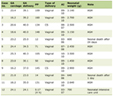 Click to view | Table 1. Clinicopathological Characteristics and Their Association With 2-Year PFS in EOC Patients |
The data also showed that 68.4% of patients did not use contraception and most patients had a normal body mass index (BMI, 50.9%). Most of the patients were stage III (60.5%). Survival decreased with increasing stage, in a clear, statistically significant trend (P < 0.001). In terms of histopathological type, the survival rate was lowest for type II tumors (21.3%) compared to type I (44.8%), and this difference was statistically significant (P = 0.008).
Most cell differentiation was G3 (87.7%), and the 2-year survival rate was 35% (P = 0.942). There was no statistical significance in PFS between lymphovascular space invasion (LVSI)-positive and LVSI-negative patients (35.5% vs. 34.9%) (P = 0.764).
All CSC markers showed that patients with low expression had higher 2-year survival than those with high expression; however, all P value over 0.05.
Receiver operating characteristic (ROC) analysis showed that none of the five CSC markers demonstrated meaningful discriminatory ability in predicting 2-year PFS (Fig. 1). The area under the curve (AUC) values ranged from 0.469 to 0.578, indicating limited discriminatory performance (Table 2). The AUC below 0.5 offers little more predictive value than random chance. CD117 was particularly underwhelming, with an AUC that actually fell below 0.5—meaning that it performed worse than chance alone. These findings were further supported by non-significant P values (P > 0.05) across all markers, confirming that none of the markers distinguished between the outcome groups.
 Click for large image | Figure 1. Receiver operating characteristic curves for CD24, CD133, CD44, CD117, and ALDH1 in predicting 2-year progression-free survival in patients with advanced epithelial ovarian cancer. ALDH: aldehyde dehydrogenase. |
Click to view | Table 2. Receiver Operating Characteristics and Youden Index |
All markers showed Youden index values below 0.2, indicating weak overall diagnostic performance. Although CD24 showed the highest Youden index (0.137) at a cut-off of 84.17, the value was too low to be meaningful for diagnostic purposes.
In summary, CD24, CD133, CD44, CD117, and ALDH1 show limited ability to distinguish outcomes and are not reliable as standalone prognostic markers in this study population.
All associations were statistically significant (P < 0.05) (Table 3). This indicates that the expression of CSC markers correlates with PFS across histopathological types. High expression of CSC markers CD24, CD133, CD44, and CD117 was correlated with poor survival. This is a sign of a more aggressive tumor and a higher risk of early recurrence. This same pattern was seen in type I and type II tumors. Type II had consistently lower survival, consistent with its more aggressive nature.
Click to view | Table 3. Association of CSC Marker Expression and Histopathological Subtypes With 1- and 2-Year PFS in Epithelial Ovarian Cancer |
For example, in CD24, 58.8% of type I patients with low expression were progression-free at 1 year, compared with 48.5% with high expression. A similar pattern was seen in type II tumors (35% vs. 18.5%). This indicates that higher CD24 expression is associated with poor prognosis in both subtypes. This trend continued for 2 years. Results for other markers were similar.
CD133 survival at 1 year was 59.6% vs. 40% in type I and 27.6% vs. 22.2% in type II. CD44 showed 63.6% vs. 51.8% in type I and 33.3% vs. 24.4% in type II. CD117 showed 57.5% vs. 48.1% in type I and 28% vs. 22.7% in type II. At 2 years, the same gap was observed, once again highlighting their ability to predict tumor aggressiveness and recurrence.
Heterogeneous survival outcomes across tumor subtypes were associated with ALDH expression. In type I tumors, high ALDH expression was associated with better survival than low expression (72.7% vs. 50%), which may indicate a favorable prognostic role. However, in type II tumors, low ALDH expression was associated with a better outcome than high ALDH expression (28.9% vs. 11.1%). These results suggest that ALDH may not have a uniform prognostic role across histological subtypes, and may reflect biological variability in tumors rather than a uniform prognostic effect. Further studies are needed to elucidate its potential context-dependent role.
To evaluate whether type I and type II categorization offers greater prognostic value than detailed histological subtypes, survival outcomes were compared between the two methodologies (Table 4). At 1 year, PFS in endometrioid cancer was 64.3%, decreasing to 46.4% at 2 years. We detected comparable trends in MC and HGSOC rates, which declined from 27.3% to 18.2% and from 25.5% to 21.3%, respectively. These patterns emphasize subtype variety and indicate inherent heterogeneity. Upon categorizing the tumors into type I and type II, a more distinct pattern emerged. At 1 year, the proportion of progression-free patients was 53.7% for type I and 25.5% for type II, declining to 44.8% and 21.3% in 2 years, respectively.
Click to view | Table 4. Histopathological Classification and PFS |
This difference persisted over time and was statistically significant (P values < 0.05). Outcomes are more effectively distinguished by classification into type I and type II rather than by specific histological subtypes. This indicates that this classification more accurately represents tumor biology and more effectively predicts PFS.
For many combinations, namely CD24–CD117, CD44–CD117, and CD24–CD44, type I tumors had higher 1- and 2-year survival rates than type II tumors. The combinations of ALDH and CD133 presented distinct and conflicting patterns. Consistent reductions in survival were observed in type II cancers in the low-expression groups of ALDH–CD44 and ALDH–CD24 during the 1- and 2-year follow-up periods. In type I cancers, lower expression of ALDH-CD117 was associated with poorer survival (47.8 vs. 56.8), and in type II cancers the opposite trend was seen, with lower expression associated with a modest increase in 1-year PFS (27.3 vs. 25). This indicates a subtype-dependent effect. The low- and high-expression groups for CD24–CD133 in type I malignancies had similar survival rates (53.3 vs. 53.8) and no significant prognostic difference; a similar trend was observed for CD44–CD133, with the low-expression group showing marginally worse survival. In type II tumors, the CD133–CD117 combination showed a temporal reversal, with lower expression associated with better 1-year survival (28.6 vs. 25) but poorer 2-year survival (14.3 vs. 22.5).
The ALDH–CD133 combination consistently showed that low expression was associated with poor survival in both type tumors at both time intervals (type I: 51.5 vs. 55.9; type II: 15 vs. 33.3). This emphasizes its unique and more reliable prognostic value compared to other marker combinations. These data support context-dependent and diverse roles for ALDH and CD133, particularly in more aggressive tumor subtypes. All dual marker combinations showed statistically significant association with survival (P < 0.05) (Table 5). In general, most combinations showed that lower expression was associated with increased probability of 1-year survival. This pattern was still evident at the 2-year follow-up, with lower survival observed, but low expression remained associated with better outcomes than high expression. Overall, type I cancers had a higher survival than type II tumors for most of the marker combinations, with significant differences in histological classifications. Taken together, these data suggest that the prognostic value of CSC markers is not an independent phenomenon but depends on the underlying tumor biology. The histopathological classification, especially the distinction between type I and type II, may serve as an effect modifier. Tumor types differ in the expression of CSC markers and in their association with survival.
Click to view | Table 5. Dual CSC Marker Expression on PFS According to Histopathological Type |
Multivariate analysis in Table 6 identified several independent predictors of PFS. First, there was a statistically significant overall effect of age (P = 0.015). The recurrence risk was 2.9-fold higher in patients aged 40–59 years than in those aged ≤ 40 years (hazard ratio (HR) = 2.914; 95% confidence interval (CI): 1.215–6.988; P = 0.017). On the other hand, patients ≥ 60 years showed no significant association with the reference group (P = 0.338).
Click to view | Table 6. Multivariate Cox Proportional Hazards Analysis of Factors Associated With Progression-Free Survival |
Tumor stage was a strongly significant predictor (P < 0.001). Patients with stage III disease had a 2.8-fold elevated risk of recurrence in comparison with patients with stage II (HR = 2.790; 95% CI: 1.506–5.168; P = 0.001). Also, the histopathological classification had a significant effect. The risk of recurrence was 1.6 times greater for type II than for type I tumors (HR = 1.633; 95% CI: 1.021–2.612; P = 0.041). However, CD44 expression was not statistically significant (P = 0.098). Patients with high CD44 expression showed a trend toward increased recurrence risk (HR = 1.896; 95% CI: 0.888–4.048), but this was not statistically significant.
In the conclusion stage, age and histopathological type are independent prognostic factors for PFS. However, CD44 expression is not an independent predictor of outcome using this model.
| Discussion | ▴Top |
Principal findings
More aggressive clinical behavior tumors, in particular type II tumors, have higher expression of CSC markers. Type II tumors are often more unstable and more prone to relapse; hence, it is not entirely surprising that this category has a stem-like cell population [1–3]. Among the markers evaluated, CD133 and ALDH were significantly more prominent than the other markers. Not much in every comparison, but enough to show a pattern, especially when looking at shorter PFS. This is consistent with previous findings where both markers are consistently related to tumor-initiating potential and treatment resistance. This shows that they may not only be markers of existence, but also markers of conduct [5, 6, 11].
Another important observation is the effectiveness of the classification systems. This dataset shows that the type I/type II paradigm is more in line with the clinical course, notably with PFS. It does not replace histology, but it may reflect a more basic feature, closer to the underlying progression of the tumor.
Clinical implications
From a therapeutic perspective, these findings have implications for a recognized but unresolved conundrum. Despite similar staging and histological features, the clinical courses of patients with EOC can be very different, sometimes within a short span. The association of CSC marker expression with lower PFS suggests that these markers might provide additional prognostic information beyond conventional parameters. Prior studies have indicated that increased expression of ALDH1 and CD133 is related to lower survival and higher recurrence rates [12, 13]. In this respect, CSC profiling may help define a subgroup of individuals with a biological predisposition to early progression, despite comparable traditional risk factors.
The importance of this is particularly evident in the first 2 years after therapy, a period when most recurrences occur. Patients with a malignancy that has a CSC-enriched profile, especially in the type II group, may need increased surveillance or a more conservative assessment of the first therapeutic response. Several studies have shown that CSC plays a role in chemoresistance and tumor repopulation after treatment [14, 15]. Another topic to consider is the use of biomarkers. The heterogeneity shown with single markers has hindered their therapeutic application. When markers are looked at together, however, the profile that emerges appears to better describe the core biology of the tumor. Recent studies reveal that stemness-related signatures or multi-marker panels may better predict differentiation than individual markers alone [16, 17]. The trends found in this analysis appear to be consistent with that methodology. Apart from prognosis these discoveries may also have important repercussions for treatment. Platinum-based chemotherapy resistance and recurrence of malignancies following an initial response have been shown to be mediated by CSC. They may stay in a relatively quiescent state and subsequently promote tumor relapse [14, 18]. Identification of CSC-enriched tumors may guide future strategies targeting this resistant cell type population.
Strengths and limitations
One of the strengths of this study is the use of a fairly homogenous cohort with EOC, minimizing the biological heterogeneity related to histology. Tumor heterogeneity has a major impact on CSC marker expression and, if not properly addressed, could complicate interpretation [4–6]. Moreover, the clinical importance is higher in advanced-stage cancer as CSC activity is significantly associated with treatment resistance and recurrence in this group [11, 16]. Immunohistochemistry, being a widely available tool in routine pathological assessment, has improved translational usefulness. However, there are some limitations that must be considered. Observational technique limits causal inference, because the association between CSC markers and development cannot be interpreted as a direct mechanical relationship. Methodological differences in immunohistochemical protocols, including scoring and cut-off criteria, may affect reproducibility, an issue that is frequently reported in CSC studies [5, 12]. Furthermore, the statistical power may be affected by the sample size and subgroup analyses, especially if several markers are examined simultaneously. The lack of extensive multivariable modeling prevents the determination of the independent prognostic relevance of CSC markers relative to established clinical factors [14, 19].
Future research directions
Future work should aim to replicate these findings in further prospective cohorts, for the inclusion into the normal clinical practice validation of CSC markers across different groups would be needed. Further studies are required to optimize the use of CSC markers. The development of composite panels or scoring systems, as opposed to focusing on single markers, may be more representative of tumor biology. Preliminary explorations of stemness-related gene signatures indicate that this strategy could boost the prediction efficiency. Integration of molecular and genomic data is a major pathway for progress. The combination of immunohistochemical CSC markers and genomic profiling may lead to better risk assessment and a more detailed understanding of tumor dynamics. Development of drugs targeting CSCs is one of the major research areas. If CSCs are indeed crucial to recurrence, strategies to eliminate them may improve long-term outcomes for individuals with OC [11, 20].
Conclusions
In this study, CSC markers alone did not independently predict PFS. While trends for worse outcomes were identified for tumors with increased expression of CSC markers, the correlations were not statistically significant in univariate or multivariate models and individual markers showed limited discriminatory efficiency. The predictive relevance of CSC markers is context-dependent and influenced by the underlying tumor biology. In the context of histopathological classification, such as type I/type II, CSC marker profiles were significantly associated with survival. This result indicates that CSC-related biological behavior may be influenced by tumor subtype instead of functioning as an autonomous prognostic predictor. Tumor stage, age, and histological classification had the greatest independent association with PFS. In several instances, the type I/type II classification produced a clearer and stronger predictive stratification than complete histologic subtyping, emphasizing its potential clinical usefulness for risk stratification.
Acknowledgments
The authors wish to extend the appreciation to Sanglah General Hospital, Dr. Cipto Mangunkusumo National General Hospital, and all respondents who have participated in this study.
Financial Disclosure
This study received allocation from Islamic Development Bank (IsDB) in alliance with Ngoerah Hospital.
Conflict of Interest
The authors declare that there is no conflict of interest.
Informed Consent
Written informed consent was obtained from all participants prior to enrollment in the study.
Author Contributions
I Nyoman Gede Budiana: study conception and design, data acquisition, data analysis, manuscript drafting, and critical revision of the manuscript. Kade Yudi Saspriyana: study conception and design, data acquisition, data analysis, manuscript drafting, and critical revision of the manuscript. Both authors read and approved the final manuscript.
Data Availability
The data supporting the findings of this study are available from the corresponding author upon reasonable request.
AI Use Declaration
The scientific content in this manuscript was not analyzed, interpreted, or created using any AI technology.
Abbreviations
ALDH: aldehyde dehydrogenase; AUC: area under the curve; BMI: body mass index; CCC: clear cell carcinoma; CD117: cluster of differentiation 117; CD133: cluster of differentiation 133; CD24: cluster of differentiation 24; CD44: cluster of differentiation 44; CSCs: cancer stem cells; EC: endometrioid carcinoma; EOC: epithelial ovarian cancer; FIGO: International Federation of Gynecology and Obstetrics; HGSOC: high-grade serous ovarian carcinoma; H-score: histochemical scoring; IHC: immunohistochemical; LGSOC: low-grade serous ovarian carcinoma; LVSI: lymphovascular space invasion; MC: mucinous carcinoma; OC: ovarian cancer; PFS: progression-free survival; RECIST: Response Evaluation Criteria in Solid Tumors; ROC: receiver operating characteristic; SPSS: Statistical Package for the Social Sciences
| References | ▴Top |
- Sung H, Ferlay J, Siegel RL, Laversanne M, Soerjomataram I, Jemal A, et al. Global cancer statistics 2022: GLOBOCAN estimates of incidence and mortality worldwide for 36 cancers in 185 countries. CA Cancer J Clin. 2024;74(1):1-35.
doi - Huang X, Li XY, Shan WL, Chen Y, Zhu Q, Xia BR. Targeted therapy and immunotherapy: Diamonds in the rough in the treatment of epithelial ovarian cancer. Front Pharmacol. 2023;14:1131342.
doi pubmed - Wang M, Zhang J, Wu Y. Tumor metabolism rewiring in epithelial ovarian cancer. J Ovarian Res. 2023;16(1):108.
doi pubmed - Fortner RT, Trewin-Nybraten CB, Paulsen T, Langseth H. Characterization of ovarian cancer survival by histotype and stage: A nationwide study in Norway. Int J Cancer. 2023;153(5):969-978.
doi pubmed - Nowicki A, Kulus M, Wieczorkiewicz M, Pienkowski W, Stefanska K, Skupin-Mrugalska P, Bryl R, et al. Ovarian cancer and cancer stem cells-cellular and molecular characteristics, signaling pathways, and usefulness as a diagnostic tool in medicine and oncology. Cancers (Basel). 2021;13(16):4178.
doi pubmed - Ma H, Tian T, Cui Z. Targeting ovarian cancer stem cells: a new way out. Stem Cell Res Ther. 2023;14(1):28.
doi pubmed - Ding J, Zhang Y, Che Y. Ovarian cancer stem cells: Critical roles in anti-tumor immunity. Front Genet. 2022;13:998220.
doi pubmed - Krolewska-Daszczynska P, Wendlocha D, Smycz-Kubanska M, Stepien S, Mielczarek-Palacz A. Cancer stem cells markers in ovarian cancer: clinical and therapeutic significance (Review). Oncol Lett. 2022;24(6):465.
doi pubmed - Ikram D, Masadah R, Nelwan BJ, Zainuddin AA, Ghaznawie M, Wahid S. CD133 Act as an essential marker in ovarian carcinogenesis. Asian Pac J Cancer Prev. 2021;22(11):3525-3531.
doi pubmed - Asian Archives of Pathology. Instructions for authors and manuscript preparation. Asian Arch Pathol. 2021;3(1). Available from: https://www.asianarchpath.com/storage/article_files/AAP_Technical_V3N1_2021.pdf.
- Fraszczak K, Barczynski B. The role of cancer stem cell markers in ovarian cancer. Cancers (Basel). 2023;16(1):40.
doi pubmed - Izycka N, Rucinski M, Andrzejewska M, Szubert S, Nowak-Markwitz E, Sterzynska K. The prognostic value of cancer stem cell markers (CSCs) Expression-ALDH1A1, CD133, CD44-for survival and long-term follow-up of ovarian cancer patients. Int J Mol Sci. 2023;24(3):2400.
doi pubmed - Izycka N, Zaborowski MP, Ciecierski L, Jaz K, Szubert S, Miedziarek C, Rezler M, et al. Cancer stem cell markers-clinical relevance and prognostic value in high-grade serous ovarian cancer (HGSOC) based on the cancer genome atlas analysis. Int J Mol Sci. 2023;24(16):12746.
doi pubmed - Luo Y, Zhang L, Liu Z, et al. Stemness index-based signature for predicting prognosis in ovarian cancer. Front Oncol. 2022;12:836916.
doi - Li L, Zhang W, Qiu J, Zhang W, Lu M, Wang J, Jin Y, et al. Stem cell-associated signatures help to predict diagnosis and prognosis in ovarian serous cystadenocarcinoma. Stem Cells Int. 2023;2023:4500561.
doi pubmed - Zhao L, Wang L, Ji W, et al. Cancer stem cells and chemoresistance in ovarian cancer. Front Oncol. 2022;12:865263.
doi - Wu Y, Wang Y, Wei M, et al. Mechanisms of cancer stem cell-mediated platinum resistance. Front Cell Dev Biol. 2021;9:693329.
doi - Yang W, Kim D, Kim DK, Choi KU, Suh DS, Kim JH. Therapeutic strategies for targeting ovarian cancer stem cells. Int J Mol Sci. 2021;22(10):5059.
doi pubmed - Wang G, Liu X, You Y, Chen S, Chang X, Yang Q. Development and clinical validation of a seven-gene signature based on tumor stem cell-related genes to predict ovarian cancer prognosis. J Ovarian Res. 2024;17(1):58.
doi pubmed - Ptak A, Rak-Mardyla A, Gregoraszczuk EL. Cancer stem cells in ovarian cancer biological role and therapeutic implications. Cells. 2022;11(5):815.
doi
This article is distributed under the terms of the Creative Commons Attribution 4.0 International License (CC BY 4.0), which permits unrestricted use, distribution, and reproduction in any medium, including commercial use, provided the original work is properly cited.
World Journal of Oncology is published by Elmer Press Inc.