World Journal of Oncology, ISSN 1920-4531 print, 1920-454X online, Open Access
Article copyright, the authors; Journal compilation copyright, World J Oncol and Elmer Press Inc
Journal website https://wjon.elmerpub.com

Original Article

Volume 17, Number 3, June 2026, pages 412-418


Could Pretreatment Computed Tomography Imaging Accurately Predict the Pathological Diagnosis of Lymph Node Involvement in Thymic Epithelial Tumors?

Figures

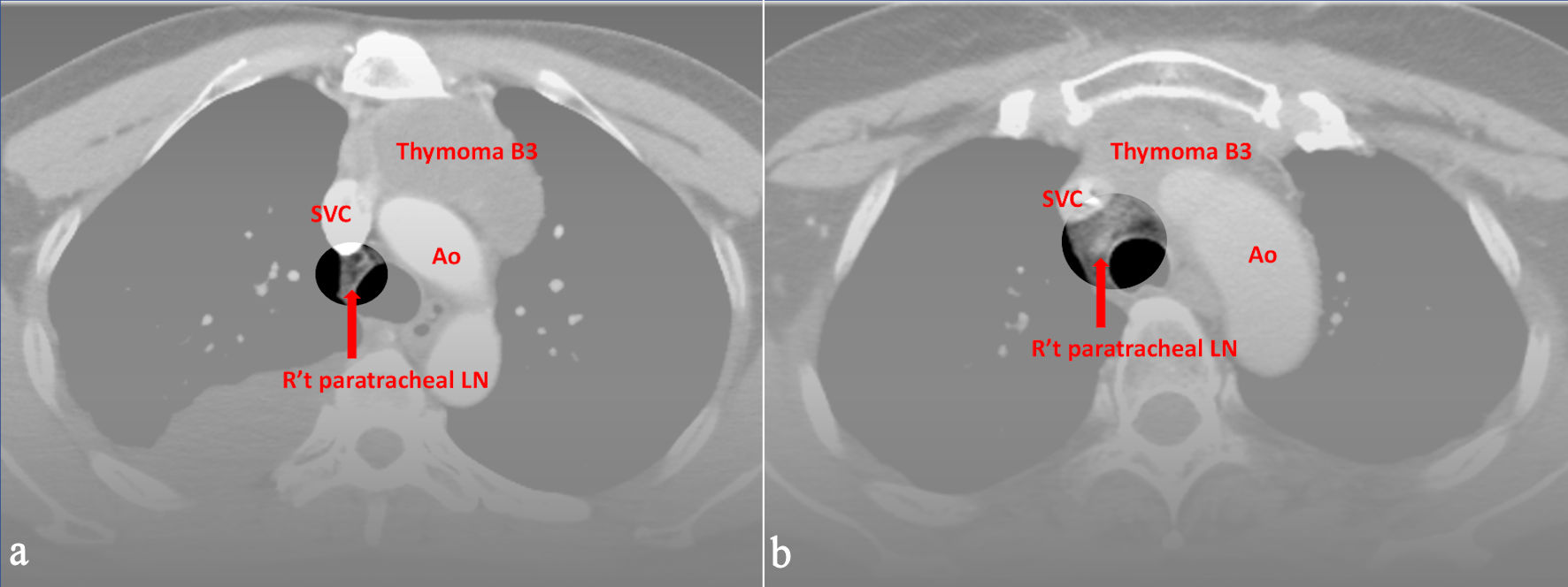
↓  Figure 1. Preoperative CT images of (a) a 52-year-old male and (b) a 49-year-old female with World Health Organization (WHO) classification B3. All three radiologists evaluated right upper paratracheal LN metastasis, and the pathological report proved the positive findings. Ao: aorta; SVC: superior vena cava; CT: computed tomography; LN: lymph node.
Figure 1.
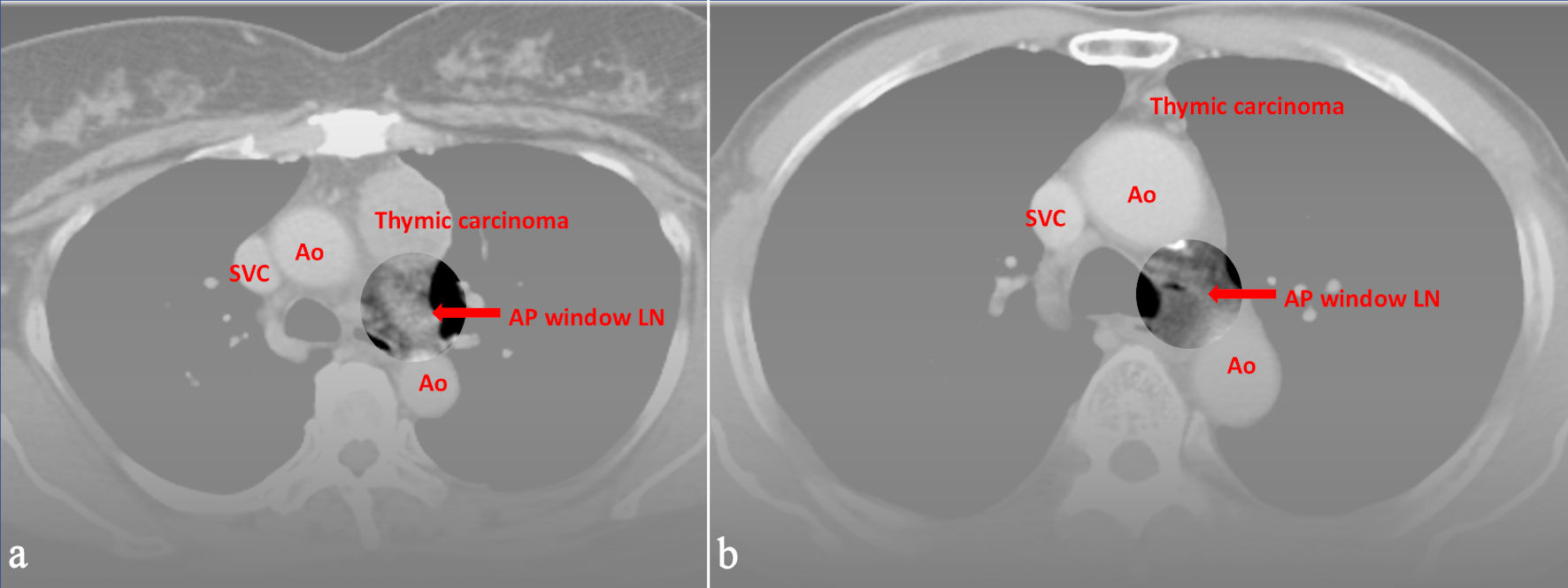
↓  Figure 2. Preoperative CT images of (a) a 59-year-old female and (b) a 74-year-old male with World Health Organization (WHO) classification thymic carcinoma. All three radiologists evaluated LN metastasis in the aortopulmonary window, and the pathological report proved the positive findings. Ao: aorta; SVC: superior vena cava; CT: computed tomography; LN: lymph node.
Figure 2.

Tables

↓  Table 1. Basic Characteristics of TETs Patients Receiving Operation and Mediastinal Lymph Node Dissection
 
Patients (n = 30)Range or %
TETs: thymic epithelial tumors; CT: computed tomography; VATS: video-assisted thoracoscopic surgery; RATS: robotic-assisted thoracic surgery; WHO: World Health Organization; M-K: Masaoka-Koga.
Gender, male (%)1343.3%
Operation age, year57.3 ± 10.938–74
Preoperative CT tumor size, cm5.6 ± 2.72–11.5
Preoperative chemotherapy or radiation310%
Operation method
  Median sternotomy930%
  Lateral thoracotomy26.7%
  VATS1446.7%
  RATS516.7%
Lymph node harvest
  N130100%
  N2310%
Mediastinal lymph node metastasis413.3%
M-K stage
  I1136.7%
  IIa413.3%
  IIb26.7%
  III723.3%
  IVa13.3%
  IVb413.3%
WHO histotype
  A + AB + B1 (low risk)1136.7%
  B2 + B3 (moderate risk)930%
  Micronodular thymoma with lymphoid stroma13.3%
  Carcinoma930%


↓  Table 2. Evaluation of Sensitivity, Specificity, and Predictive Values by Identifying TETs Lymph Node Metastasis From CT Image
 
TPFPTNFNSensitivitySpecificityPPVNPVAccuracy
Mediastinal lymph node metastasis was recorded in four patients with TETs in the pathology reports. TETs: thymic epithelial tumors; CT: computed tomography; TP: true positive; FP: false positive; TN: true negative; FN: false negative; PPV: positive predictive value; NPV: negative predictive value.
TETs (n = 30)
  Radiologist A42240100%92.3%66.7%100%93.3%
  Radiologist B410160100%61.5%28.6%100%66.7%
  Radiologist C46200100%76.9%40%100%80%


↓  Table 3. Previous Studies for TETs LN Metastasis
 
Years of inclusionLymph node harvest patient numbersLymph node metastasis rate
TETs: thymic epithelial tumors; LN: lymph node; Thy: thymoma; TC: thymic carcinoma; NETT: neuroendocrine thymic tumor.
Kondo et al, 2003, [10]1990–1994Thy: 1,0641.8%
TC: 18326.8%
Park et al, 2013 [11]1995–2010TC: 2623.1%
Weissferdt et al, 2012 [12]1985–2011TC: 3036.7%
Weksler et al, 2015 [13]1988–2009Thy: 44213.3%
Weksler et al, 2015 [14]1988–2011TC: 17664.1%
NETT: 5335.9%
Hwang et al, 2016 [15]1996–2011Thy: 995.1%
TC: 3225.0%
Fang et al, 2018 [16]2014–2016Thy: 2432.1%
TC: 2425.0%
NETT: 850.0%
Current study2008–2023Thy: 219.5%
TC: 922.2%