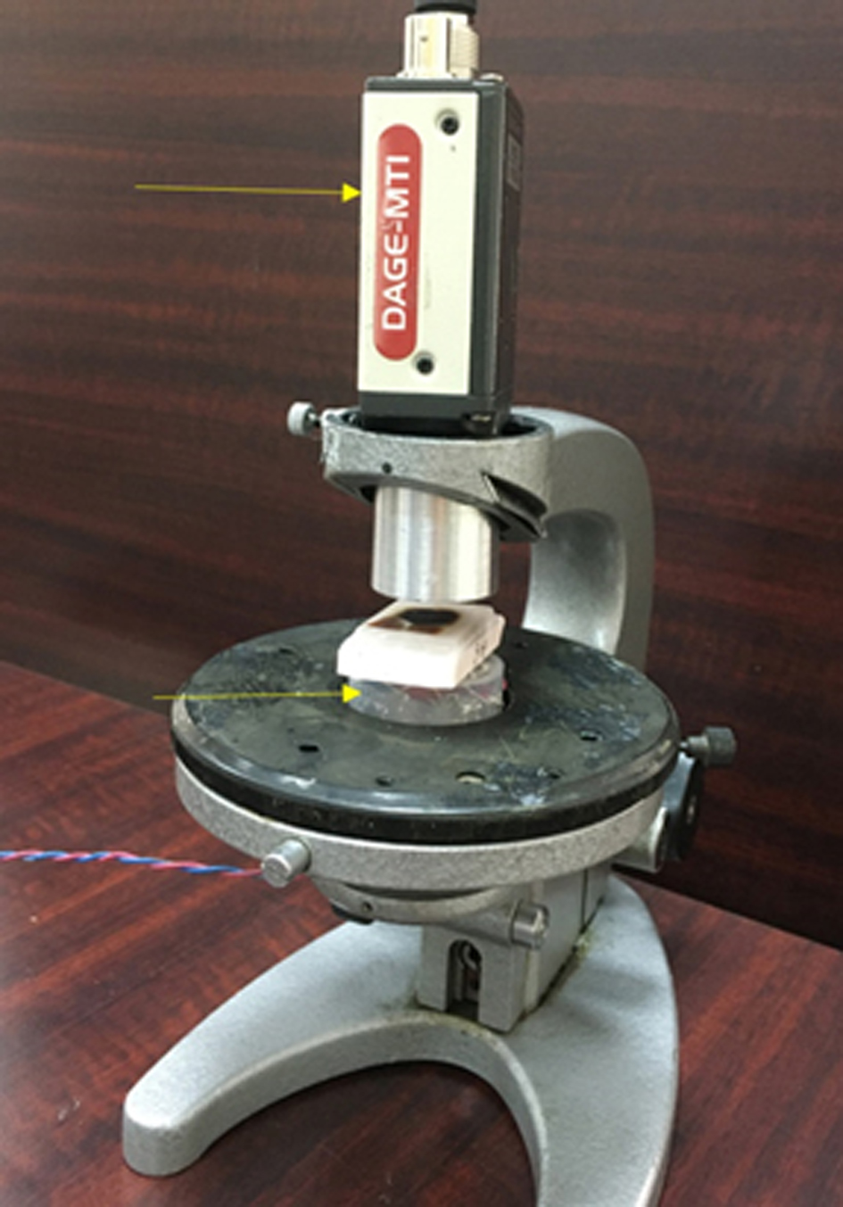
↓ Figure 1. Setup of the experiment. Arrows are
used to show the CCD camera and LEDs. Between them lies biological tissue. To prevent the influence of
daylight, experiments were conducted in complete darkness.
| World Journal of Oncology, ISSN 1920-4531 print, 1920-454X online, Open Access |
| Article copyright, the authors; Journal compilation copyright, World J Oncol and Elmer Press Inc |
| Journal website https://wjon.elmerpub.com |
Original Article
Volume 16, Number 3, June 2025, pages 311-316
Differentiating Malignant and Healthy Areas in Isolated Kidney Samples Through Infrared Visualization Techniques
Figures
Table
| Kidney tumor | 32 cases |
| Tumor dimensions, cm | 3.5 - 7 |
| Age, years | 46 - 76 (mean 59.5) |
| Gender | |
| Female | 13 |
| Male | 19 |
| Histomorphological types | |
| Clear cells | 16 |
| Papillary | 6 |
| Chromophobe | 7 |
| Collecting duct cancer | 3 |
| Stage | |
| T1a N0M0 tumor ≤ 4 cm | 14 |
| T1b N0M0 tumor > 4 cm, but ≤ 7 cm | 18 |