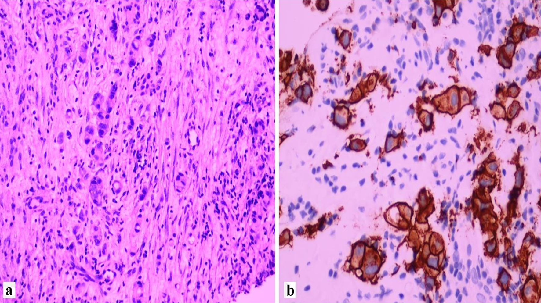
↓ Figure 1. Histological features of
HER2-positive invasive lobular carcinoma. HER2: human epidermal growth factor receptor 2.
| World Journal of Oncology, ISSN 1920-4531 print, 1920-454X online, Open Access |
| Article copyright, the authors; Journal compilation copyright, World J Oncol and Elmer Press Inc |
| Journal website https://wjon.elmerpub.com |
Original Article
Volume 16, Number 4, August 2025, pages 357-364
Clinicopathological Features of HER2 Expressing Lobular Carcinoma of Breast
Figures
Tables
| Demographics | Patients/specimens (n = 28) |
|---|---|
| HER2: human epidermal growth factor receptor 2; ILC: invasive lobular carcinoma. | |
| Median age (years) | 54 |
| Stage at presentation (TNM) | |
| Tumor (T) | |
| T1 | 2 (7.1%) |
| T2 | 6 (21.4%) |
| T3 | 7 (25%) |
| Metastatic | 8 (28.5%) |
| Unknown | 5 (17.8%) |
| Node (N) | |
| Nx | 5 (17.9%) |
| N0 | 1 (3.6%) |
| N1 | 1 (3.6%) |
| N2 | 2 (7.1%) |
| N3 | 19 (67.9%) |
| Treatment | |
| Neoadjuvant chemotherapy with anti-HER2 | 11 (39.2%) |
| Adjuvant chemotherapy with anti-HER2 | 7 (25%) |
| Palliative | 8 (28.6%) |
| Unknown | 2 (7.1%) |
| Status | HER2-negative p-ILC | HER2-overexpressing ILC | Total |
|---|---|---|---|
| Data are expressed as N (Chi-square). HER2: human epidermal growth factor receptor 2; ILC: invasive lobular carcinoma; p-ILC: pleomorphic invasive lobular carcinoma. | |||
| Alive without disease | 8 (0.18) | 7 (0.15) | 15 |
| Alive with advanced disease | 2 (0.21) | 4 (0.18) | 6 |
| Dead | 7 (0.02) | 9 (0.01) | 16 |
| Total | 17 | 20 | 37 (grand total) |
| P-value | 0.689 | ||