Figures
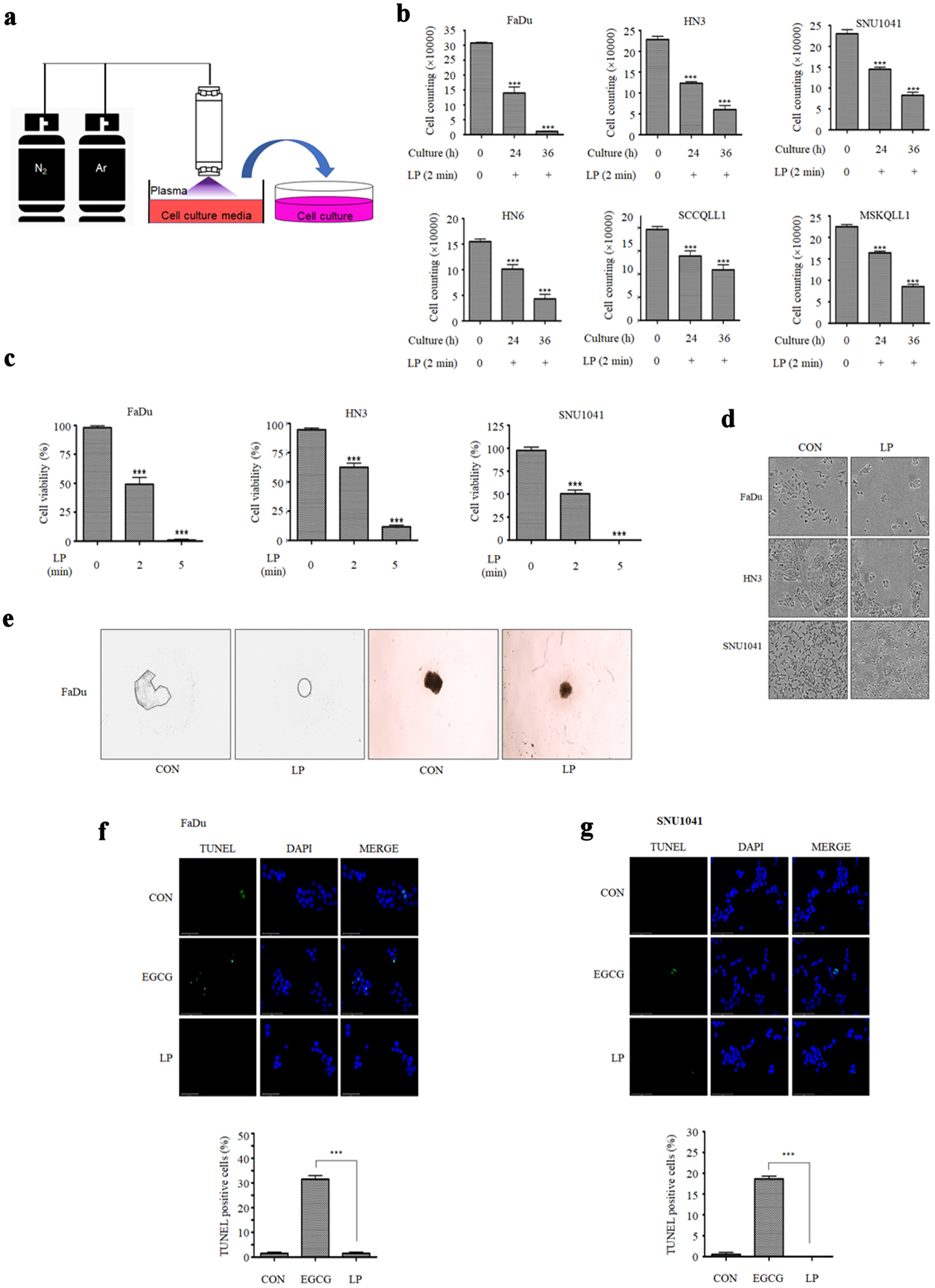
↓ Figure 1. Liquid plasma (LP) induces cell death
in HNC cells. (a) N2 gas was set to 200 standard cubic centimeters per minute (sccm) and Ar
gas to 1,500 sccm. Cell culture medium was treated with N2/Ar plasma for 2 min per
milliliter. (b) HNC cells were treated with LP for 24 or 36 h, stained with trypan blue, and then number
of live cells was measured. (c) FaDu, HN3, and SNU1041 cells were treated with LP (N2/Ar
plasma-treated medium for 2 min or 5 min per milliliter), and the viability was measured using CCK-8
assays after 24 h. (d) The morphology of cells was observed under a microscope 24 h after LP treatment.
(e) A 3D spheroid culture of FaDu cells was observed following LP treatment. (f) FaDu cells and (g)
SNU1041 cells were subjected to TUNEL assays (scale bar: 50 µm). *P < 0.05, **P < 0.01, ***P
< 0.001. HNC: head and neck cancer; CCK: Cell Counting Kit; 3D: three-dimensional.
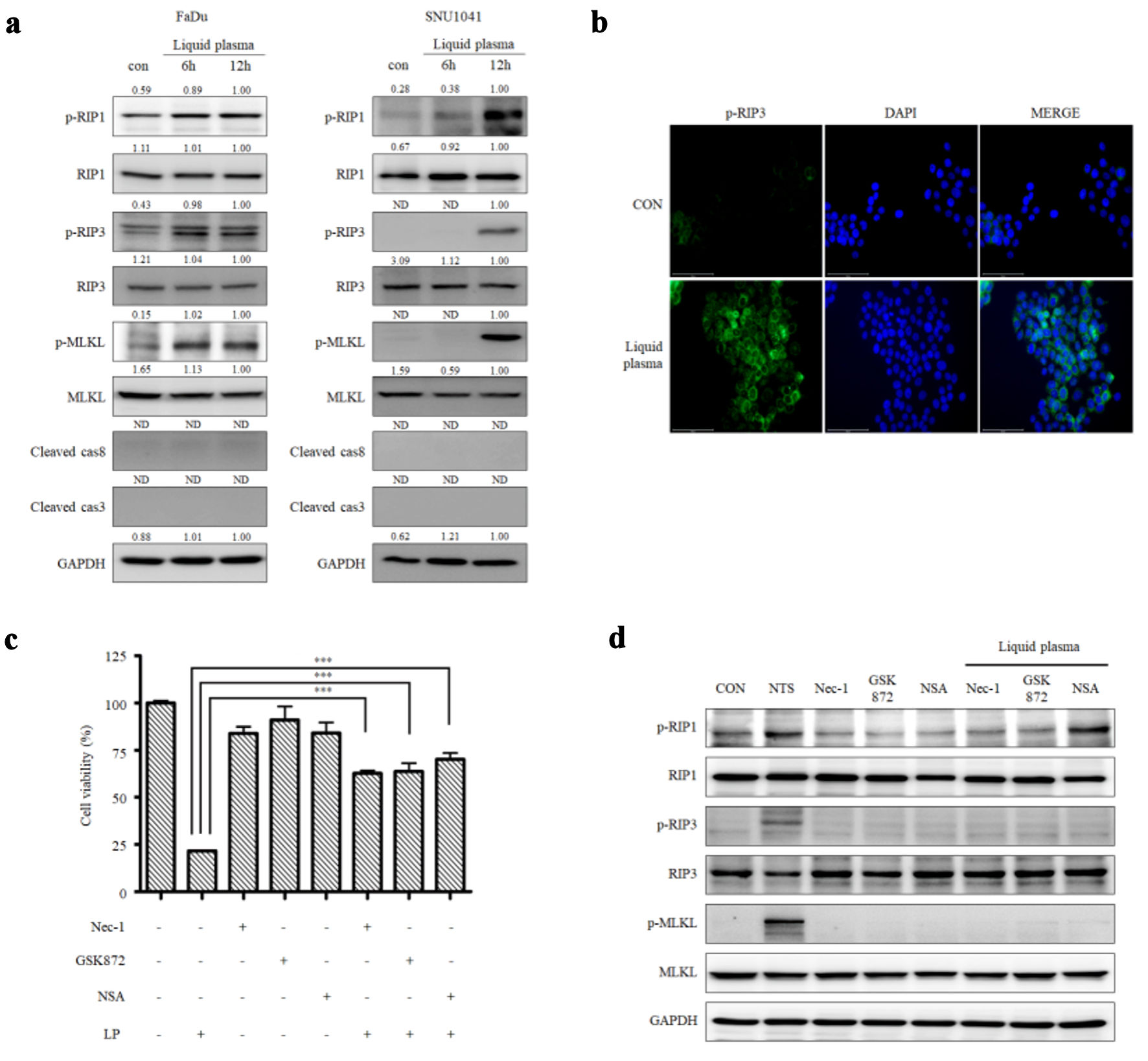
↓ Figure 2. Liquid plasma (LP) induces
necroptosis in FaDu and SNU1041 cells. (a) FaDu and SNU1041 cells were treated with LP for 6 or 12 h,
and the expression levels of p-RIP1, RIP3, p-RIP1, RIP, p-MLKL, MLKL, cleaved caspase-8, cleaved
caspase-3, and GAPDH were analyzed by Western blotting. (b) FaDu cells were treated with LP for 6 h, and
p-RIP3 expression was visualized using fluorescence staining (scale bar: 50 µm). (c) FaDu cells
were treated with LP, a p-RIP1 inhibitor (Nec-1), a p-RIP3 inhibitor (GSK872), and a p-MLKL inhibitor
(NSA), and cell viability was measured using CCK-8 assays. (d) The expression levels of p-RIP1, RIP3,
p-RIP1, RIP, p-MLKL, MLKL, and GAPDH were analyzed by Western blotting. *P < 0.05, **P < 0.01,
***P < 0.001. RIP: receptor-interacting protein; Nec-1: necrostatin-1; MLKL: mixed lineage kinase
domain-like protein; GAPDH: glyceraldehyde-3-phosphate dehydrogenase; NSA: necrosulfonamide; CCK: Cell
Counting Kit.
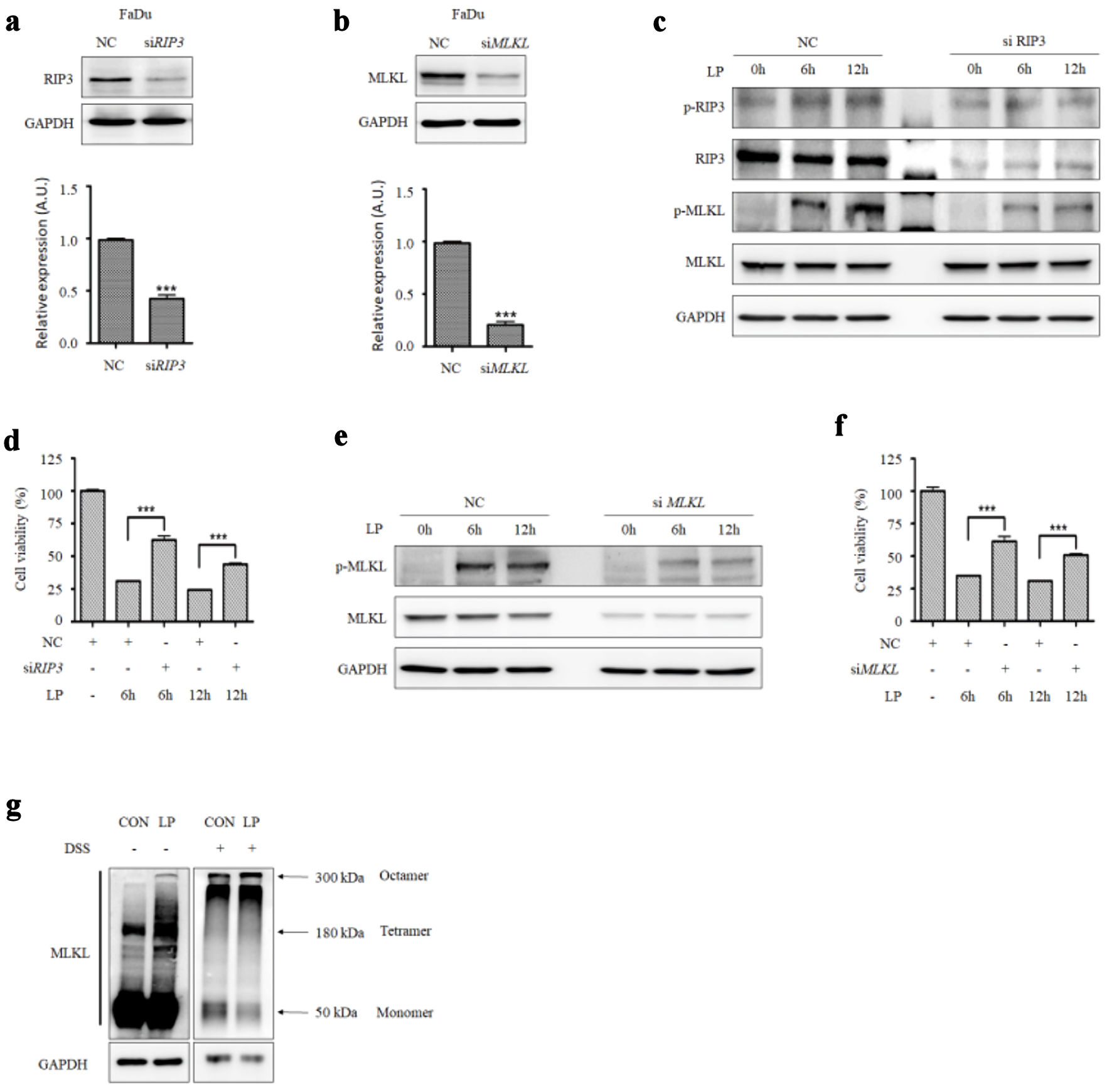
↓ Figure 3. RIP3 and MLKL levels affect the
sensitivity to liquid plasma (LP) in HNC cells. (a) RIP3 and (b) MLKL were knocked down in FaDu cells
using siRNA. (c) FaDu cells transfected with siRIP3 RNA and (e) siMLKL RNA were treated with LP for 6 or
12 h, and protein expression levels (RIP3, MLKL, and their phosphorylated forms) were analyzed by
Western blotting. (d) FaDu cells transfected with siRIP3 RNA and (f) siMLKL RNA were treated with LP for
6 or 12 h, and cell viability was measured using Cell Counting Kit (CCK-8) assays. (g) MLKL
oligomerization was detected by Western blotting under non-reducing conditions. *P < 0.05, **P <
0.01, ***P < 0.001. HNC: head and neck cancer; RIP: receptor-interacting protein; MLKL: mixed lineage
kinase domain-like protein; GAPDH: glyceraldehyde-3-phosphate dehydrogenase.
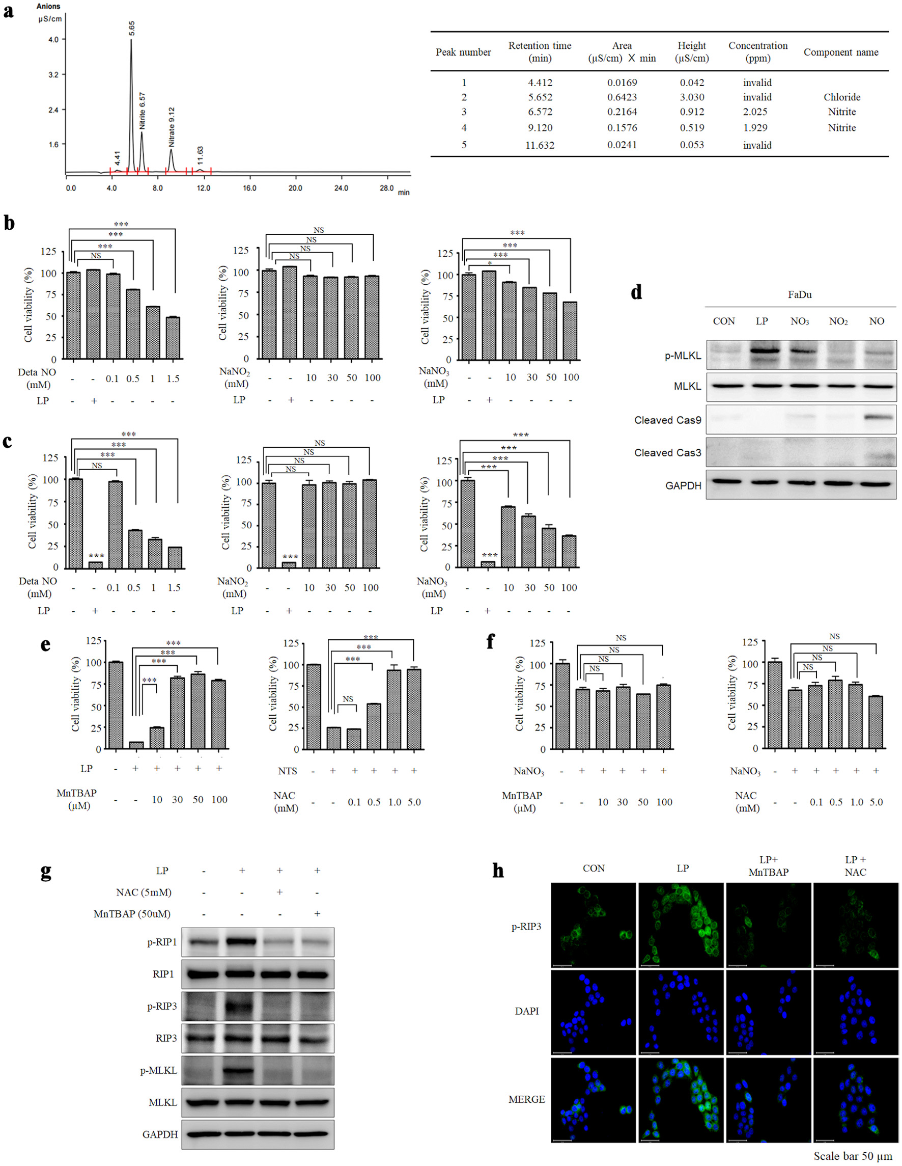
↓ Figure 4. Peroxynitrite in liquid plasma (LP)
induces necroptosis in HNC cells. (a) The components of LP were analyzed using a chromatographic ion
analyzer. (b) Human dermal fibroblasts (HDF) and (c) FaDu cells were treated with DetaNO,
NaNO2, NaNO3, and LP, representing nitrite and nitrate, which are presumed
components of plasma. (d) FaDu cells were treated with DetaNO, NaNO2, NaNO3, and
LP, and the expression levels of p-MLKL, MLKL, cleaved caspase-9, cleaved caspase-3, and GAPDH were
analyzed by Western blotting. (e) FaDu cells were treated with LP, and the peroxynitrite inhibitor
MnTBAP was added at different concentrations. Cell viability was measured using CCK-8 assays. (f) FaDu
cells were treated with NaNO3, and the peroxynitrite inhibitor MnTBAP was added at different
concentrations. Cell viability was measured using CCK-8 assays. (g) FaDu cells were treated with NAC and
MnTBAP for 12 h, and the expression levels of p-RIP2, RIP1, p-RIP3, RIP3, p-MLKL, MLKL, and GAPDH were
analyzed by Western blotting. (h) The expression of p-RIP3 following LP treatment was confirmed using
fluorescence staining (scale bar: 50 µm). *P < 0.05, **P < 0.01, ***P < 0.001. HNC: head
and neck cancer; MLKL: mixed lineage kinase domain-like protein; MnTBAP: manganese (III) tetrakis
(4-benzoic acid) porphyrin chloride; RIP: receptor-interacting protein; GAPDH:
glyceraldehyde-3-phosphate dehydrogenase; DAPI: 4′,6-diamidino-2-phenylindole; NS:
non-significant.
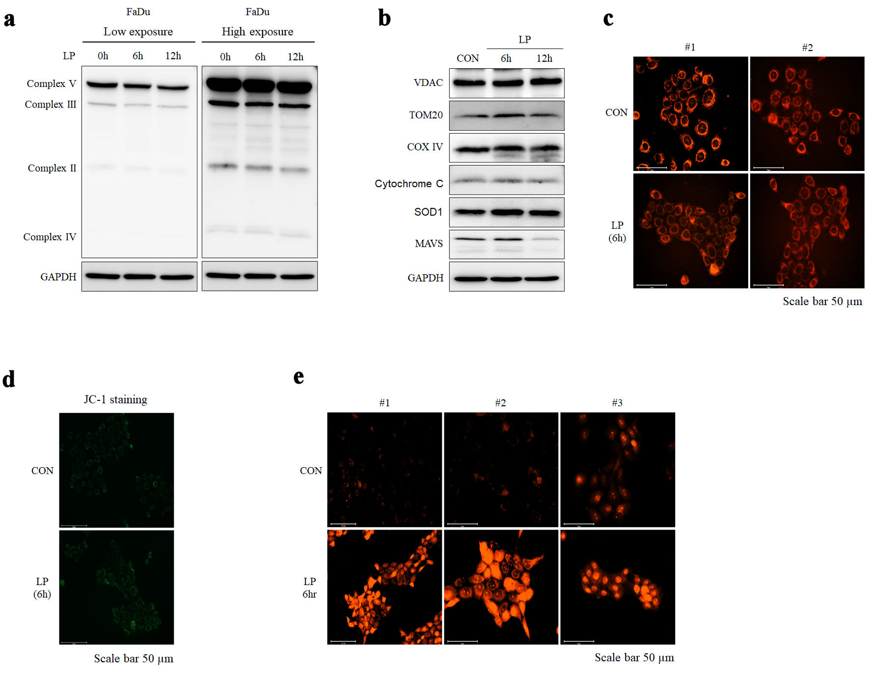
↓ Figure 5. Liquid plasma (LP) treatment induces
mitochondrial stress in HNC cells. (a, b) The expression of mitochondria complex and mitochondrial
proteins (VADC, TOM20, COX IV, cytochrome c, SOD, MAVS) following LP treatment was confirmed using
Western blotting. (c, d) Changes in mitochondrial morphology after LP treatment were observed using
fluorescence staining with MitoRed and JC-1, respectively (scale bar: 50 µm). (e) ROS levels
following LP treatment were assessed using fluorescence staining with MitoSox (scale bar: 50 µm).
HNC: head and neck cancer; MAVS: mitochondrial antiviral signaling; GAPDH: glyceraldehyde-3-phosphate
dehydrogenase; ROS: reactive oxygen species.
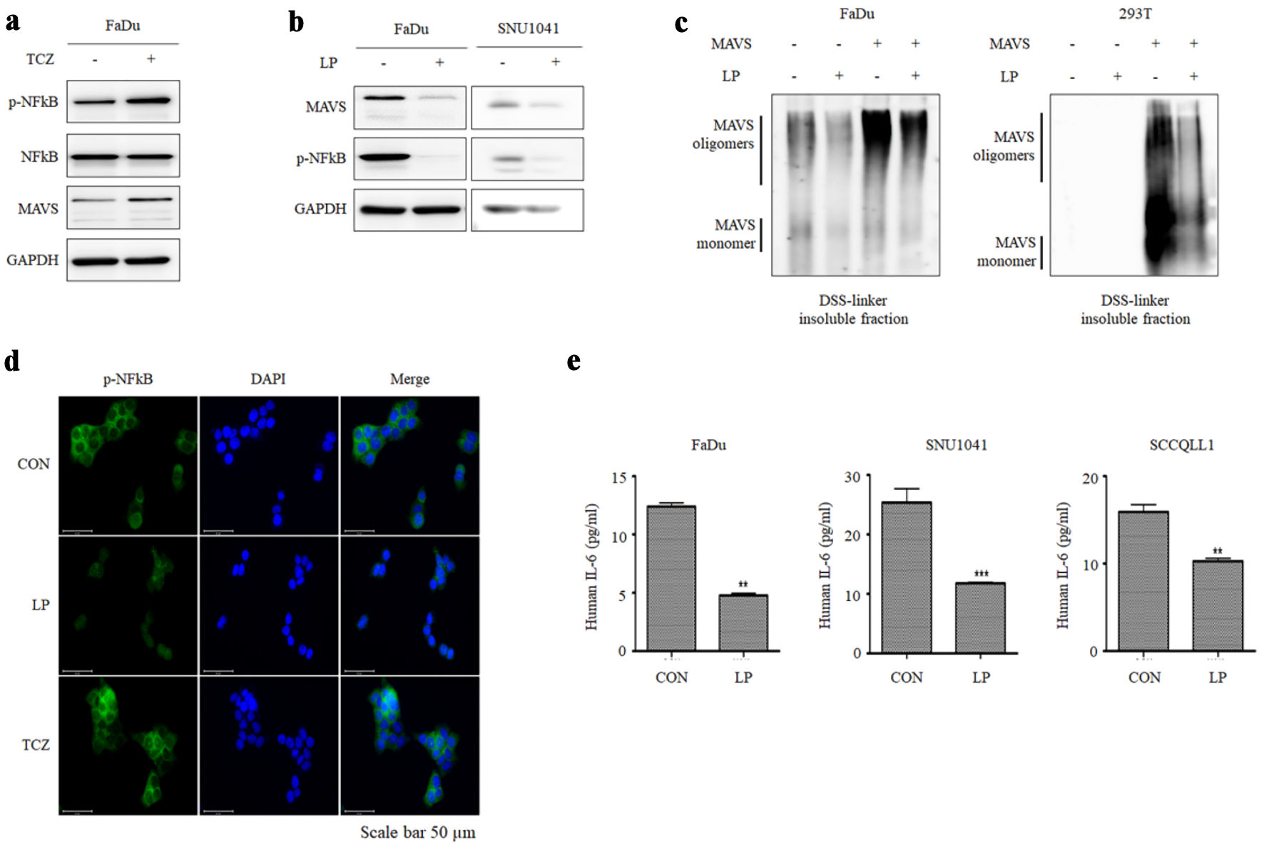
↓ Figure 6. Liquid plasma (LP) inhibits
necroptosis-mediated inflammation by degrading MAVS aggregates. (a) FaDu cells were treated with TCZ (30
µg/mL TNF-α, 300 µM CHX, and 10 µM zVAD-fmk) for 12 h, and the expression of MAVS
and p-NF-κB was observed. (b) LP treatment suppressed the expression of MAVS and p-NF-κB in
FaDu and SNU1041 cells. (c) After inducing MAVS oligomerization in FaDu and 293T cells, the degradation
of oligomerized MAVS by LP treatment was observed. (d) FaDu cells treated with both LP and TCZ, changes
in NF-κB expression levels and nuclear translocation were analyzed using fluorescence microscopy
(scale bar: 50 µm). (e) LP treatment inhibited TCZ-induced IL-6 production in FaDu and SNU1041
cells. IL-6 levels in the supernatant were measured using ELISA assay. **P < 0.01, ***P < 0.001.
MAVS: mitochondrial antiviral signaling; TNF-α: tumor necrosis factor alpha; CHX: cycloheximide;
NF-κB: nuclear factor kappa B; IL: interleukin; GAPDH: glyceraldehyde-3-phosphate dehydrogenase;
DAPI: 4′,6-diamidino-2-phenylindole; TCZ: TNF-α + cycloheximide + zVAD-fmk.