Figures
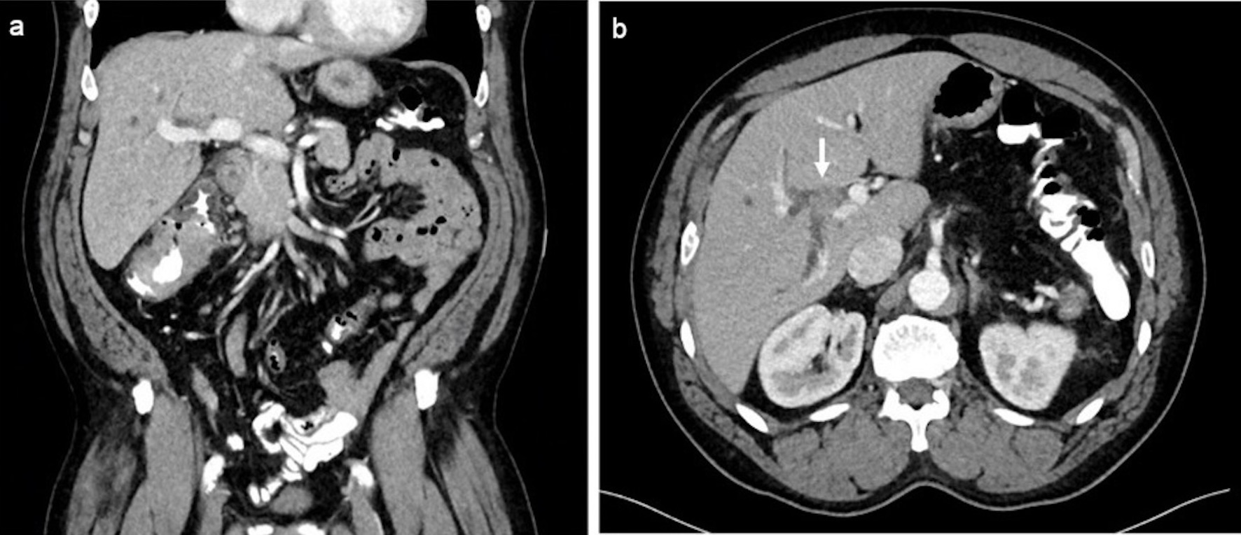
↓ Figure 1. (a) Coronal post-contrast-enhanced
computed tomography (CT) demonstrating mass-like wall thickening of the cecum spanning approximately 8
cm in length compatible with biopsy-proven adenocarcinoma. (b) Axial post-contrast-enhanced portal
venous phase CT showing an enhancing mass within the right bile duct (arrow) measuring 1.1 × 1.3 cm
with associated right-sided intrahepatic biliary dilation.
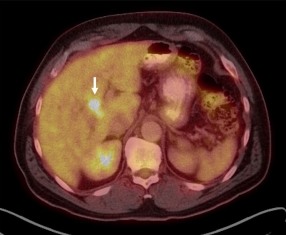
↓ Figure 2. Fused axial 18F-FDG PET/CT showing
intense, focal FDG uptake (SUV max 9.6) in a 2.1 cm lesion near the porta hepatis (arrow). 18F-FDG:
18F-fluorodeoxyglucose; CT: computed tomography; PET: positron emission tomography; SUV: standardized
uptake value.
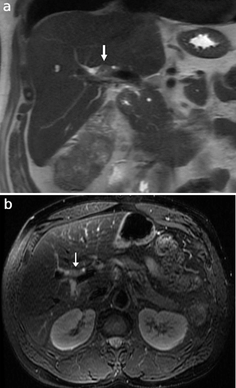
↓ Figure 3. Representative (a) coronal T2 HASTE
sequence and (b) axial T2 fat saturated sequence MRCP images showing an intermediate T2 signal mass
(arrows) within the proximal right intrahepatic bile duct corresponding to the focus of radiotracer
uptake on prior PET. HASTE: half-Fourier acquired single-shot turbo spin-echo; MRCP: magnetic resonance
cholangiopancreatography; PET: positron emission tomography.
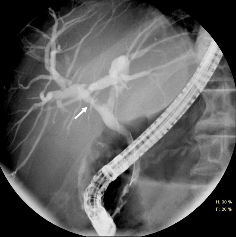
↓ Figure 4. ERCP cholangiogram showing irregular
filling defect within the right bile duct corresponding to the location of the known mass (arrow). ERCP:
endoscopic retrograde cholangiopancreatography.
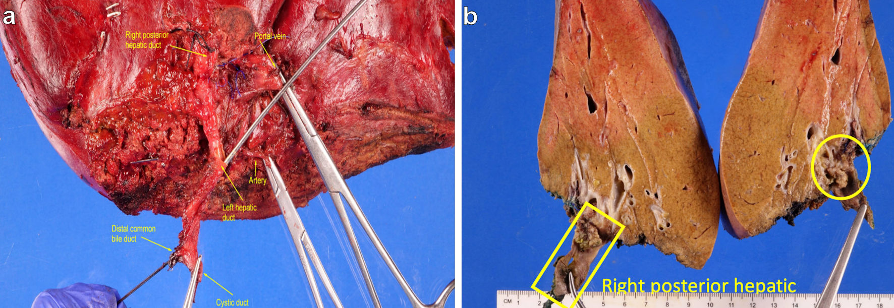
↓ Figure 5. (a) Hepatic hilar region. (b) Yellow
square marks right posterior hepatic bile duct and yellow circle marks tan-yellow friable and exophytic
mass involving the duct.
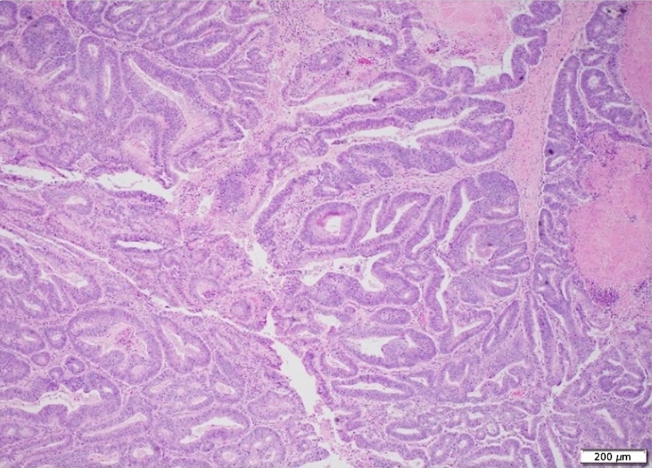
↓ Figure 6. Section of the cecal mass with
invasive moderately differentiated adenocarcinoma
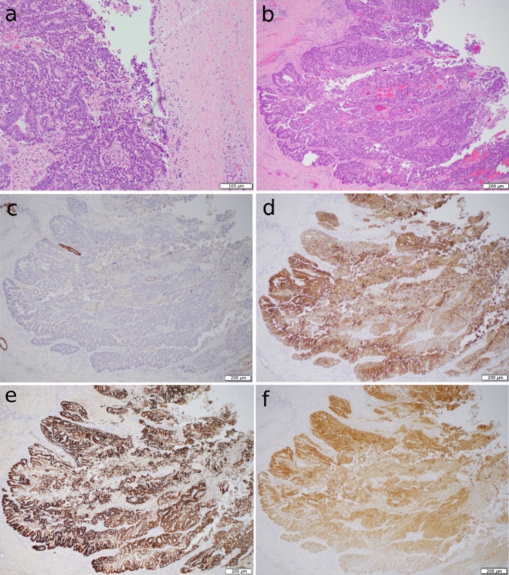
↓ Figure 7. (a, b) Sections of the right hepatic
bile duct with exophytic mass. The mass has similar morphology to the cecal mass. (c) CK7
immunohistochemistry with no expression. (d, e, f) CK20, CDX2 and SATB2 immunohistochemistry with
positive diffused expression, respectively. CK7: cytokeratin 7; CK20: cytokeratin 20.
Table
↓ Table 1. Immunohistochemical and Molecular Profiles of Resected Cecal
and Bile Duct Lesions
|
|
Cecal mass |
Bile
duct lesion |
| MMR: mismatch repair; MSI: microsatellite instability; CK: cytokeratin. |
| Immunophenotype |
|
CK7 negative |
|
|
CK20 positive, patchy |
|
|
CDX2 positive, strong and diffuse |
|
|
SATB2 positive, strong and diffuse |
| Molecular profile |
MMR intact |
MMR intact |
|
MSI absent |
MSI absent |
|
BRAF wild-type |
KRAS G12D point mutation |
|
KRAS G12D point mutation |
APC K1245* point mutation |
|
|
APC E1408* point mutation |
|
|
PIK3CA E545K point mutation |
|
|
PPP2R1A C148Lfs* 26 point mutation |
|
|
ZFHX3 R2203C point mutation |