Figures
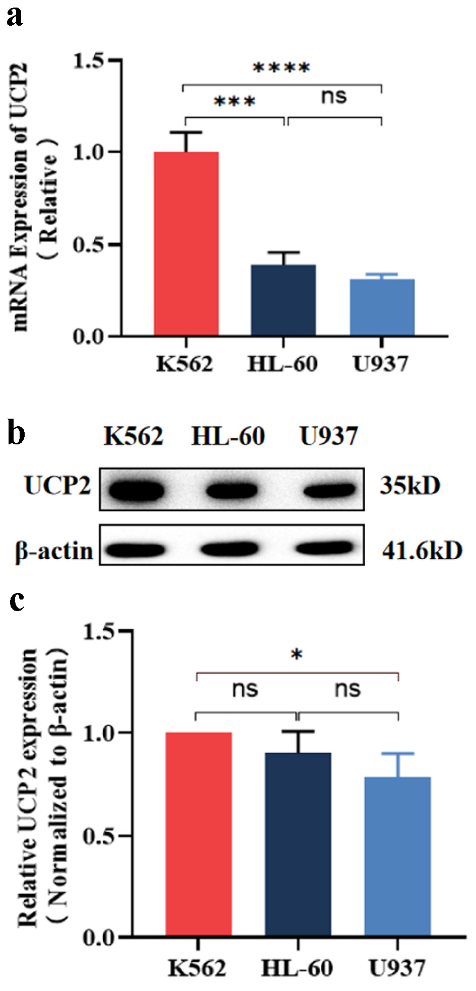
↓ Figure 1. Expression of UCP2 in ML cell lines.
(a) Expression levels of UCP2 mRNA in K562, HL-60, and U937. Western blot bands (b) analysis of UCP2
protein expression levels (c) in K562, HL-60, and U937. Data were presented as mean ± SD of the
relative changes (n = 3). *P < 0.05, **P < 0.01, ***P < 0.001, ****P < 0.0001, ns: no
significant P > 0.05. ML: myeloid leukemia; UCP2: uncoupling protein 2; SD: standard deviation.
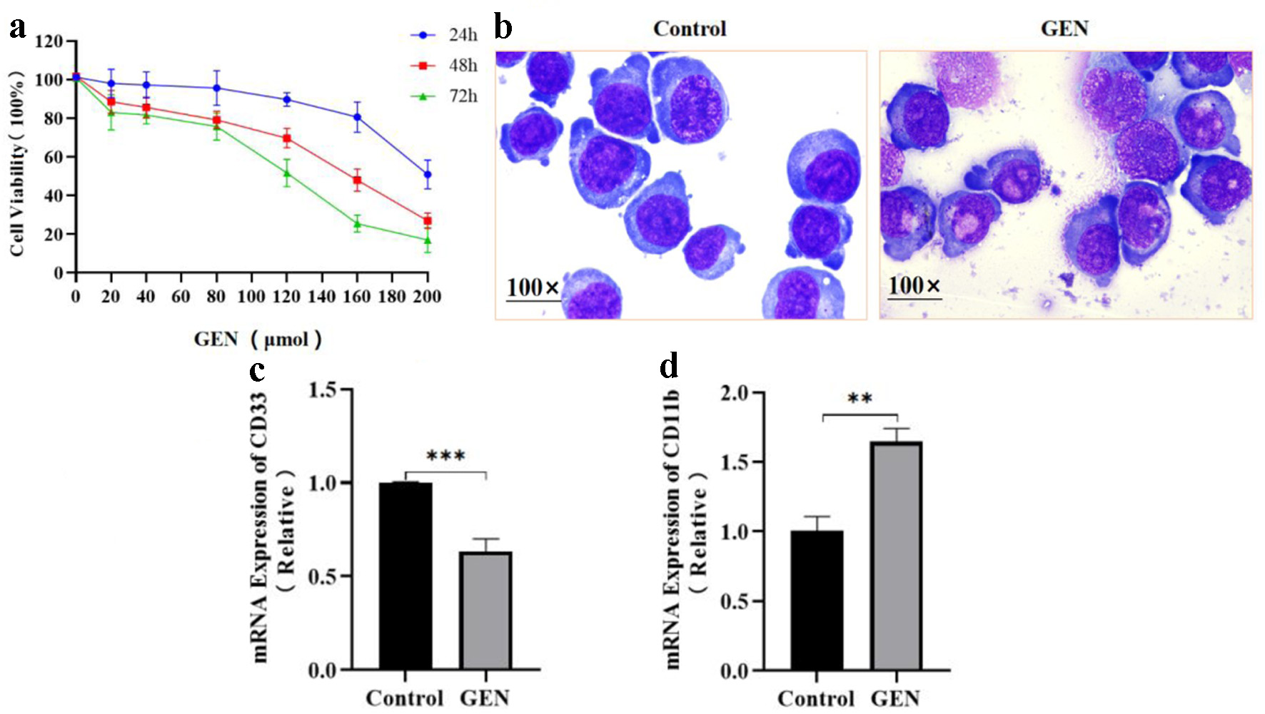
↓ Figure 2. The effect of GEN on the
proliferation and differentiation of K562 cells. (a) Cell proliferation was evaluated after treatment
with various concentrations of GEN (0, 20, 40, 80, 120, 160, and 200 µmol/L) in K562 cells,
utilizing the CCK-8 assay. (b) Cell morphology was observed through Wright-Giemsa staining. The mRNA
expression levels of CD33 (c) and CD11b (d) were assessed in K562 cells treated with 112 µmol/L GEN
for 72 h. Data were presented as mean ± SD of the relative changes (n = 3). **P < 0.01,
***P < 0.001. CCK-8: cell counting kit-8; GEN: genipin; SD: standard deviation.
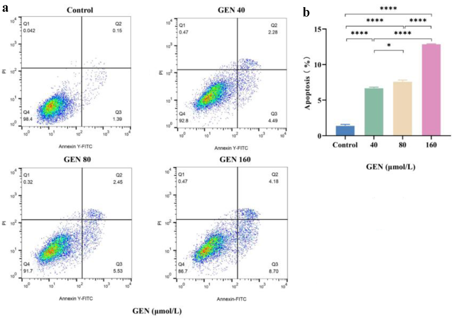
↓ Figure 3. The promoting effect of GEN on
apoptosis of K562 cells. (a) Flow cytometry analysis was conducted to assess the promotion of apoptosis
in K562 cells after 72 h of treatment with 40, 80, and 160 µmol/L of GEN. (b) Column analysis of
the promotion of apoptosis in K562 cells after 72 h of treatment with 40, 80, and 160 µmol/L of
GEN. All data were presented as mean ± SD of the relative changes (n = 3). *P < 0.05, **P <
0.01, ***P < 0.001, ****P < 0.0001. GEN: genipin; SD: standard deviation.
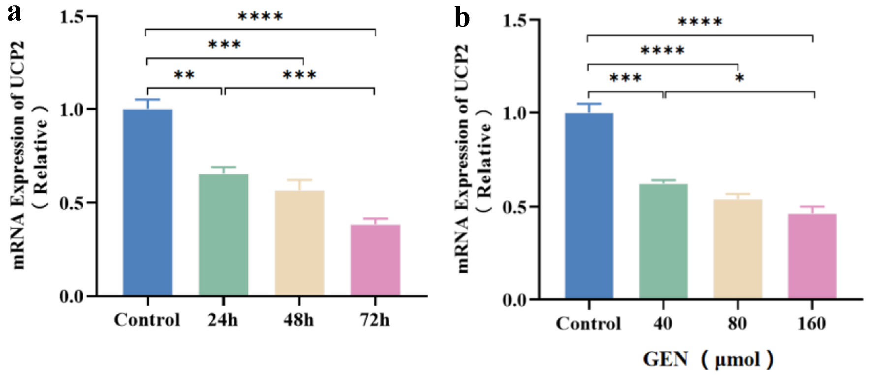
↓ Figure 4. The expression level of UCP2 mRNA in
K562 cells is inhibited by GEN. Relative expression levels of UCP2 mRNA in K562 cells treated with 160
µmol/L GEN at 24, 48, and 72 h (a). Relative expression levels of UCP2 mRNA in K562 cells treated
with GEN at different concentrations (40, 80, and 160 µmol/L) for 72 h (B). All data were presented
as mean ± SD of the relative changes (n = 3). *P < 0.05, **P < 0.01, ***P < 0.001,
****P < 0.0001. GEN: genipin; SD: standard deviation; UCP2: uncoupling protein 2.
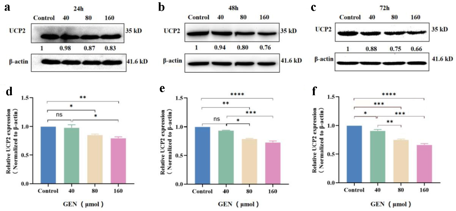
↓ Figure 5. The expression level of UCP2 protein
in K562 cells was inhibited in a concentration-dependent manner. Western blot analysis of UCP2 protein
expression levels in K562 cells treated with various concentrations of GEN (40, 80, and 160 µmol/L)
for 24 h (a), 48 h (b), and 72 h (c). Grayscale analysis bar chart depicting the expression levels of
UCP2 protein in K562 cells treated with GEN at different concentrations for 24 h (d), 48 h (e), and 72 h
(f). All data were presented as mean ± SD of the relative changes (n = 3). *P < 0.05, **P <
0.01, ***P < 0.001, ****P < 0.0001, ns: no significant P > 0.05. GEN: genipin; SD: standard
deviation; UCP2: uncoupling protein 2.
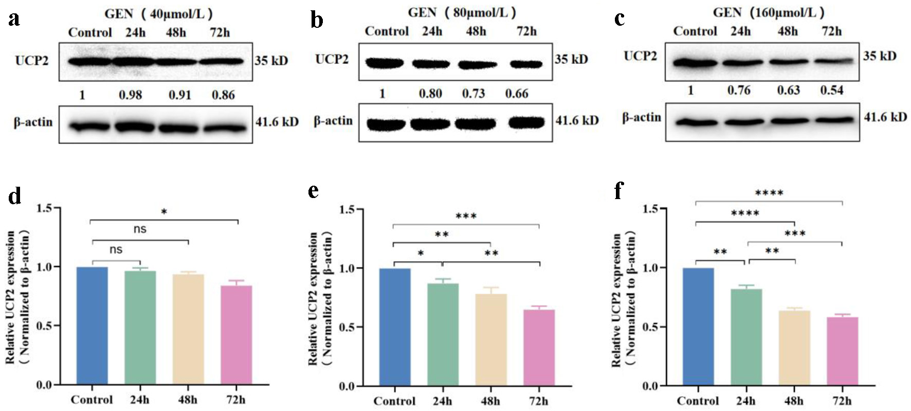
↓ Figure 6. The expression level of UCP2 protein
in K562 cells was inhibited in a time-dependent manner. Western blot analysis indicated that GEN at
concentrations of 40 µmol/L (a), 80 µmol/L (b), and 160 µmol/L (c) suppressed UCP2
protein expression levels in K562 cells at 24, 48, and 72 h. Grayscale analysis bar chart of GEN at 40
µmol/L (d), 80 µmol/L (e), and 160 µmol/L (f) inhibiting UCP2 protein expression levels
in K562 cells at 24, 48, and 72 h. All data were presented as mean ± SD of the relative changes (n
= 3). *P < 0.05, **P < 0.01, ***P < 0.001, ****P < 0.0001, ns, no significant P > 0.05.
GEN: genipin; SD: standard deviation; UCP2: uncoupling protein 2.
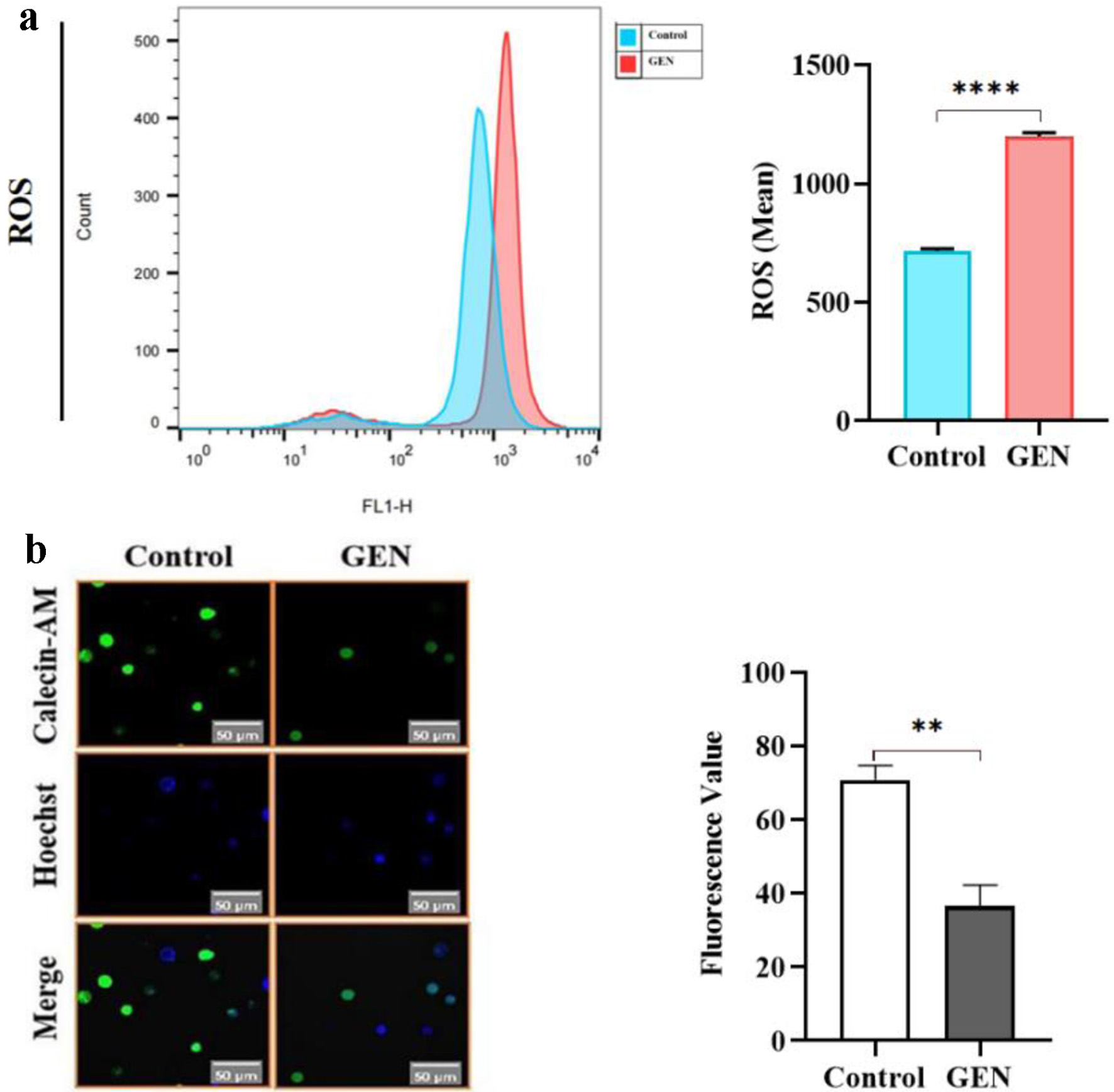
↓ Figure 7. GEN induces mitochondrial damage in
K562 cells. (a) Promotion of ROS generation by GEN in K562 cells. (b) Promotion of MPTP by GEN in K562
cells. All data were presented as mean ± SD of the relative changes (n = 3). **P < 0.01, ****P
< 0.0001. GEN: genipin; MPTP: membrane permeability transition pore; ROS: reactive oxygen species;
SD: standard deviation.
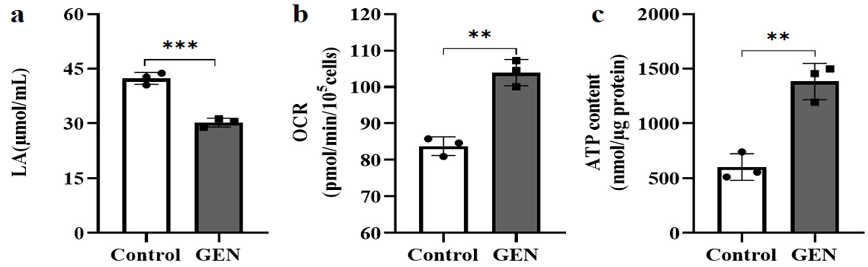
↓ Figure 8. The effect of GEN on the energy
metabolism pathway of K562 cells. (a) GEN inhibits LA production. (b) GEN increases oxygen consumption.
(c) GEN promotes ATP production. The control group was cultured under normal conditions. The
experimental group was treated with 112 µmol/L GEN. All data were presented as mean ± SD of
the relative changes (n = 3). **P < 0.01, ***P < 0.001. ATP: adenosine triphosphate; GEN: genipin;
SD: standard deviation.
Tables
↓ Table 1. General Clinical Data in ML Patients
|
Groups |
N
|
M/F
|
Age
range (years) |
| AML: acute myeloid leukemia; CML: chronic myeloid leukemia; ML: myeloid leukemia. |
| AML |
25 |
11/14 |
36 - 81 |
| CML |
22 |
15/7 |
30 - 78 |
| Control |
10 |
6/4 |
44 - 77 |
↓ Table 2. The PCR Primer Sequence
|
Primer name |
The
primer sequence (5′-3′) |
| PCR: polymerase chain reaction; UCP2: uncoupling protein 2. |
| UCP2-F |
CCCAAAGGCAGAAGTGAAG |
| UCP2-R |
CCCAATGTTGCTCGTAATG |
| CD33-F |
CCCAGCTCTCTGTGCATGTGA |
| CD33-R |
GAGTGCCAGGGATGAGGATTT |
| CD11b-F |
ACTTGCAGTGAGAACACGTATG |
| CD11b-R |
TCATCCGCCGAAAGTCATGTG |
| GAPDH-F |
CAATGACCCCTTCATTGACC |
| GAPDH-R |
GACAAGCTTCCCGTTCTCAG |
↓ Table 3. The Expression of UCP2 mRNA in ML Patients
|
Groups |
N
|
UCP2
(high/low cases) |
| AML: acute myeloid leukemia; CML: chronic myeloid leukemia; ML: myeloid leukemia; UCP2:
uncoupling protein 2. |
| AML |
25 |
14/7 |
| CML |
22 |
14/5 |